Download
1 / 40
400 likes | 595 Views
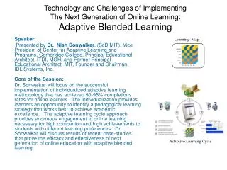
Speaker: Kumar Saurabh. Uveitis: Definition. * International Uveitis Study Group Definition.

E N D
Uveitis: Definition * International Uveitis Study Group Definition Group of diseases characterized by severe sight threatening intraocular inflammation primarily involving the uveal tract which extend occasionally to the contiguous structures like vitreous, optic nerve head, cornea and sclera.*
Problem Statement Prevalence: 0.73% worldwide Age group: 20-50 years Five to 20% of legal blindness Tedious diagnostic workup Elusive aetiologies Permanent structural damage Low therapeutic index of medications
Aetiology of Uveitis Idiopathic Uveitis: 35.7% Uveitis Secondary to Systemic Disease Infectious Uveitis Lens Induced Uveitis Masquerade Syndrome
Goals of Uveitis Management Reduction of visual morbidity Proper identification of the specific disease Cost effective utilization of resources
Approach: Ways to achieve that History Clinical Assessment: Site, stage and vision Differential Diagnosis Tailored Laboratory Tests Correlation with Ancillary Tests Definitive Treatment Control of Adverse Effects
History: Salient Points Detailed and Suggestive Geographic History Family History Demography: Age, gender, race Personal History: Pets, diet, drugs, sexual Systemic Diseases Ocular History
Ocular History Onset: Insidious or Sudden Laterality: Unilateral or Bilateral Severity: Mild or Severe Pattern: Single or Multiple Episodes Duration: Acute or Chronic Treatment
Clinical Assessment: Ocular Visual Acuity External : Lids, lacrimal glands Conjunctiva & Sclera: Congestion, nodules Cornea: Keratitis, ulceration, keratopathy Keratic precipitates Anterior chamber: Flare and cells Iris: Surface, nodules, new vessels Vitreous: Haze, detachment, hemorrhage Retina: Retinitis, new vessels, oedema Choroid: Choroiditis, neovascularization
Keratic Precipitates Mutton fat keratic precipitates in a case of granulomatous uveitis
Keratic Precipitates Fine keratic precipitates in a case of non-granulomatous uveitis
Keratic Precipitates Old keratic precipitates seen in the Arlt’s triangle of corneal endothelium
Keratic Precipitates Large keratic precipitates seen in case of granulomatous uveitis
Anterior Chamber Reaction Flare * 0 : Absence + : Barely detectable ++ : Moderate, iris details visible +++ : Marked, iris details not visible ++++: Intense, fixed fribrinous aqueous Cell * 0 : Nil 0.5 + : 1-5 cells + : 6-15 cells ++ : 16-25 cells +++ : 26-50 cells ++++ : >50 cells *Standardization of Uveitis Nomenclature (SUN). AJO 2005;140: 509-16
Anterior Chamber Reaction Cells and flare in uveitic eyes
Vitreous HazeDirect Ophthalmoscope 0 : Clear vitreous + : Few, normal view ++ : Moderate scattered opacities, obscured view +++ : Many opacities, blurring of view ++++ : Dense opacities, no view
Vitreous HazeIndirect Ophthalmoscope ++++ : Optic nerve head obscured +++ : Optic nerve head visible, blurred border ++ : Better view of retinal blood vessels + : Better view of retinal blood vessels & ONH + : Blurring of nerve fiber layer striations 0 : Nerve fiber layer well defined
Retinal Features Retinitis Primary vasculitis Secondary vasculitis Retinal detachment Macular oedema Neovascularization
Choroidal Features Active choroiditis Inactive choroiditis Choroidal granulomas Choroidal neovascularization Choroidal vasculitis
Site of Uveitis Anterior Uveitis: Iritis, iridocyclitis, Anterior cyclitis Intermediate Uveitis: Posterior cyclitis, Hyalites, Basal Retinochoroiditis Posterior Uveitis: Chorioretinitis, Retinochoroiditis, Neuroretinitis, Choroiditis
Systemic Examination Mucocutaneous system • Erythema nodosum • Oral ulcerations • Kaposi sarcoma • Kearatoderama • Circinate balanitis • Vitiligo • Sarcoid granulomas Musculoskeletal system • Ankylosing spondylitis • Rheumatoid arthritis • Psoriasis • Behcet’s disease • Reiter’s syndrome
Differential Diagnosis Working diagnosis Basis for laboratory investigation Basis for treatment
Laboratory Investigations • To rule out infective etiology • To alleviate risks of treatment • To find out systemic disease • To find out etiology of masquerade syndrome • To come to specific diagnosis • Academic purposes Aims
Laboratory Investigations • Granulomatous uveitis • Recurrent uveitis (>3 attacks) • Uveitis in a child • Positive leads on examination • Posterior uveitis • Retinal vasculitis • Worsening on steroids Indications
Laboratory Investigations Complete Blood Count Rheumatoid factor (RA) Antinuclear Antibody (ANA) Anti ds-DNA Antibody Anti Neutrophil Cytoplasmic Antibody (ANCA) Angiotensin Converting Enzyme C- reactive Protein X-ray Chest and Spine Toxoplasma, Rubella, Cytomegalovirus, Herpes simplex (TORCH) Test Mantoux Test HLA Typing
Ancillary Investigations Ultrasonography: Media opacity Fluorescein Angiography: Macular oedema, Chorioretinitis, Disc leakage, Response Vitreous Biopsy: Lymphoma, Endophthalmitis Chorioretinal Biopsy:
Specific Therapy Ocular Toxoplasmosis Regimen 2. Clindamycin 300mg, 4 times Daily, 4-6 weeks PLUS Sulphadiazine: 2 gm, D1 1 gm four times daily, 4-6wks PLUS Prednisolone: 40-60 mg/ day From D3-D7, tapered Regimen 1. Pyrimethamine: 75mg, D1 25mg/day, 4-6 weeks PLUS Sulphadiazine: 2 gm, D1 1 gm four times daily, 4-6wks PLUS Prednisolone: 40-60 mg/ day From D3-D7, tapered PLUS Folinic Acid: 5mg twice weekly
Specific Therapy Ocular Toxocariasis Peripheral Lesion: Follow up Posterior Pole Involvement: Prednisolone 40mg/day and Thiobendazole 20mg/day. 5-7 days Vitrectomy
Nonspecific Therapy: Medical Steroids: Topical, periocular, systemic Indications: Active Inflammation Prevention and treatment of complications Infiltration of retina, choroid and optic nerve
Nonspecific Therapy: Medical Nonsteroidal Antinflammatory Drugs Indication: To maintain lower dose of topical steroids
Nonspecific Therapy: Medical Immunosuppressive Therapy Vision threatening intraocular inflammation Inadequate response to steroids Serious steroid induced side effects Contraindication of steroid therapy
Nonspecific Therapy: Medical • Immunosuppressive Therapy Absolute Indications: Behcet’s disease Sympathetic Ophthalmia Vogt-Koyanagi-Harada Syndrome Wegener’s Granulomatosis Polyarteritis Nodosa
Nonspecific Therapy: Medical LASER and Cryotherapy Indications: LASER for choroidal neovascularization Cryotherapy for refractory pars planitis
Nonspecific Therapy: Surgical Pars plana Vitrectomy Indications: Pars planitis Subretinal neovscular membrane
Treatment Response* *Standardization of Uveitis Nomenclature (SUN). AJO 2005;140: 509-16 Inactive Uveitis : Rare cell in anterior chamber Improvement : Two step decrease in cells Reduction in cell count to Grade 0 Worsening : Two step increase in cells Increase in cell count from 3+ to 4+ Steroid resistance : Two weeks Immunosuppressive resistance: Three months
Treatment of Complications Management of cataract Management of glaucoma Management of band shaped keratopathy and other corneal complications