Download
1 / 45
460 likes | 1.05k Views
Presented May 2003. Resuscitation Of Newborn. Darko J. Vodopich MD Anesthesia Resident @ CWRU - MHMC. Revised by: Greg Gordon MD. Perinatal stress. Cathecolamines (CCA) are good - if neonate is deprived CCA less survival rate CCA maintain CO

E N D
Presented May 2003 Resuscitation Of Newborn Darko J. Vodopich MD Anesthesia Resident @ CWRU - MHMC Revised by: Greg Gordon MD
Perinatal stress Cathecolamines (CCA) are good - if neonate is deprived CCA less survival rate CCA maintain CO CCA redistribute blood flow towards important areas BP and HR MVO2 Neonates with CCA have higher Apgar scores CCA are important in a transition to extrauterine life
Perinatal Cardiorespiratory physiology Fetal lungs 24 days -arises from the foregut 26-28 weeks -terminal airways developed 30-32 weeks -final surface active material (SAM) developed Plasma ultrafiltrate is a normal part of the lungs Every day IU (intrauterine) 50-150 ml/kg/day of plasma is produced Plasma is swallowed in the gut and excreted by kidneys
Perinatal Cardiorespiratory physiology Plasma ultrafiltrate (2) 2/3 is expelled during vaginal delivery 1/3 is removed capillaries, lymphatics, breathing If fluid is retained into lungs causes TTN(transient tachypnea of newborn). Causes: Small infants Preterm infants Rapidly born Cesarean section born babies
Perinatal Cardiorespiratory physiology Normal breathing - 30/min @ 90 sec of age
Perinatal Cardiorespiratory physiology Normal breathing - 30/min @ 90 sec of age (reminder) Normal breathing - 40-60/min @ few minutes of age: Removal of increased CO2 produced by high metabolic rate Helps maintain FRC
Perinatal Cardiorespiratory physiology Circulation of the fetus: RV ~ 2/3 of CO LV ~ 1/3 CO Foramen ovale Ductus arteriosus Blood is coming from placenta - high O2 content 95% of the blood coming from placenta goes to LA through foramen ovale
Circulation of the fetus: The numbers are combined ventricular output
Perinatal Cardiorespiratory physiology Circulation of the newborn: PVR is due to pulmonary expansion, breathing, pH, and O2 tension If neonate is born by CS - PAP’s and PVR PVR is : Hypoxia Acidosis Hypovolemia Hypoventilation Atelectasis Cold
Perinatal Cardiorespiratory physiology Changes in circulation of the newborn: PVR - pulmonary blood flow Right/left shunting will be decreased LA pressures are , and seal foramen ovale Ductus arteriosus closes (10-14 days) in response to O2 Ach Parasympathetic nerve stimulation PG If PaO2 ~ 60-100 = closes, if 300-500 = remains open
Asphyxia PaO2 PCO2 pH Uteroplacental blood flow Maternal or Fetal disease (cause)
Asphyxia Intrauterine asphyxia: PaO2 decreases from 25-40 to 5 mmHg Anaerobic metabolism occurs pH drops < 7.0 : respiratory and metabolic acidosis Lactate is accumulating in the body Redistribution of blood flow in the body CO starting normal is now decreasing Because of high doses of opioids in the blood fetus may survive severe hypoxia (may reduce total O2 consumption)
Asphyxia Intrauterine asphyxia: (monkeys)
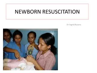
Initial evaluation of Newborn Bulb syringe suction Dry towel Breathing stimulation Strip the blood from umbilical cord: Increase blood volume Increase respiratory rate Increase lung water PAP PaCO2 Be aware that baby can be hypovolemic from early clamping of the umbilical cord
Initial evaluation of Newborn Prolonged suction may cause vomiting and arrhythmias Examine HEENT Choanal atresia NG tube suction Cor Pulmo Abdomen Extremities Do regular Apgar scores: 1 and 5 minutes
Disorders associated with Asphyxia • Maternal conditions • Elderly primigravida (>35 years of age) • Diabetes • Hypertension • Toxemia
Disorders associated with Asphyxia • Maternal treatment with any of the following: • Glucocorticoids • Diuretics • Antimetabolites • Reserpine, lithium • Magnesium • Ethyl alcohol • -Adrenergic drugs (to stop premature labor) • Abnormal estriol levels
Disorders associated with Asphyxia • Anemia (hemoglobin level less than 10 g/100 mL) • Blood type or group isoimmunization • Previous birth of child with a hereditary disease • Current maternal infection or infection during pregnancy with rubella, herpes simplex, or syphilis • Abruptio placentae • Placenta previa • Antepartum hemorrhage • History of previous infant with • jaundice, • thrombocytopenia • cardiorespiratory distress • congenital anomalies
Disorders associated with Asphyxia • Narcotic, barbiturate, tranquilizer, or psychedelic drugs • Ethyl alcohol intoxication • History of previous neonatal death • Prolonged rupture of membranes
Disorders associated with Asphyxia Conditions of labor and delivery • Forceps delivery other than low elective • Vacuum extraction delivery • Breech presentation or other abnormal presentation • Cesarean section • Prolonged labor • Prolapsed umbilical cord • Cephalopelvic disproportion • Maternal hypotension • Sedative or analgesic drugs given intravenously within 1 hour of delivery or intramuscularly within 2 hours of delivery
Disorders associated with Asphyxia • Fetal conditions • Multiple births • Polyhydramnios • Meconium-stained amniotic fluid • Abnormal heart rate or rhythm • Acidosis (fetal scalp capillary blood) • Decreased rate of growth (uterine size) • Premature delivery • Amniotic fluid surfactant test negative or intermediate within 24 hours of delivery
Disorders associated with Asphyxia • Neonatal conditions • Birth asphyxia • Birth weight (inappropriate for gestational age) • Meconium-staining of the skin, nails, or umbilical cord • Signs of cardiorespiratory distress
Apgar Scores • 8 - 10: • 90% of all newborn • Suction the baby • Be sure is normothermic • Be sure to repeat Apgar @ 5 minutes
Apgar Scores • 5 - 7: • Mild asphyxia suffered • Usually respond to vigorous stimulation • If not ventilate 80-100% face mask • Score should go up
Apgar Scores • 3 - 4: • Moderately depression @ birth • Usually they are cyanotic, bag/mask ventilation • Place ET tube if necessary • Draw ABG from umbilical artery (leave in) • Use medication based on ABG’s
Apgar Scores • 0 - 2: • Severe asphyxia • Immediate resuscitation • Pulmonary resuscitation • Vascular resuscitation (see next slides for resuscitation)
Pulmonary resuscitation • Breath for the baby 30-60/min • Hold every 5th breath for 2-3 sec for atelectasis • Apply 1-3 cm H2O of PEEP • Respect 5 differences in airway compared to adults • Closely monitor effects of ventilation • Keep POX ~ mid 90’s, and try wean off high inspired FiO2 - retinopathy of prematurity • Provide routine tracheal suctioning • 1st tracheal intubation apply suction and leave 2nd in place if necessary
Pulmonary resuscitation • Be aware of meconium aspiration • If baby < 2000 g rare meconium aspiration • 10% meconium mother stain fluid 60% babies will have in the trachea • 15% will develop respiratory complications • 10% will have PTX or pneumomediastinum • All neonates should be observed for 24 h • Prone to Persistent Fetal Circulation syndrome • Unless severe asphyxia at birth good prognosis
Pneumothorax • In 1% all vaginal deliveries • In 10% meconium stained deliveries • 2-3% neonates require mechanical ventilation • To suspect:always look the chest movement • To diagnose: • CXR • Small clod light placed over the thorax (glow) • Rx: 22-G blunt needle 2nd intercostal space
Surfactant: • Greatly improved outcome in premature neonates • Decreased incidence of: • Pulmonary gas leaks • Hyaline membrane disease • BPD • Pulmonary interstitial emphysema • Rx: 5ml/kg intratracheal after delivery • Be aware of initial transient desaturation
Vascular resuscitation: Umbilical Artery Catheterization • 3.5 F if weight < 1500 g • 5 F if weight > 1500 g • Advance 3-5 cm from the umbilical cord cut • Carefully: 0.1 ml of air may obstruct the blood flow to the legs for several hours The oximetric system for measuring arterial oxygen saturation continuously. The catheter contains fiberoptics that transmit light to and from blood passing the catheter tip
Vascular resuscitation: Umbilical Venous Catheterization • Insert ~ 3-5 cm into IVC (see size on the slide before) • Connect the catheter into transducer • Proof to be in IVC - deflection during inspiration • Be aware that air bubbles may cross foramen ovale and emboli brain
Correction of Acidosis: • Respiratory acidosis increase ventilation • Metabolic acidosis HCO3- • If using Tromethamine* be aware of: • Hypoglycemia • Hypocalcemia • Hypokalemia • Apnea * THAM is a 0.3 molar solution of tris(hydroxymethyl)- aminomethane adjusted to a pH of approximately 8.6 with 0.5% acetic acid. The solution is hypertonic (380 milliOsmoles/liter). Administer by slow IV push.
Correction of Acidosis: Potential side effects with administration of HCO3- • HCO3- are hypertonic ~1800 mOsm/L rapid intravascular infusion intracranial hemorrhage • H+ + HCO3- CO2 to increase 1-3 mmol/L if ventilation is inadequate cardiac arrest • In acidotic, hypovolemic neonates with intense peripheral vasoconstriction hypotension may occur correcting the acidosis ( PVR the neonate will not have adequate blood volume to fill intravascular space.
Correction of Acidosis: Who requires HCO3-: • If Apgar 2 min ~, 5 min ~ 5 and controlled ventilation and tactile stimulation 2 ml/kg HCO3- • pH < 7.00 and PaCO2 is < 35 mmHg 1/ of base deficit* should be corrected * Base Deficit = 0.5 x weight in kg x (24 - [HCO3-])
Correction of Hypovolemia: • ~ 60 % of neonates are hypovolemic early clamping of umbilical cord • Detection of hypovolemia: • ABP • Physical exam: • Skin color • Perfusion • Capillary refill time • Pulse volume • Extremity temperature • CVP < 4 mmHg
Rx. of Hypovolemia: • 10 mL/kg of LR • 1-2 g/kg of 25% albumen • 10 mL/kg of plasma • Cross-matched blood • O-negative blood
Other causes of Hypotension: • We must consider other causes of hypotension: • Alcohol • Hypomagnesemia • Hypocalcemia • Polycythemia (increased PVR)
Cardiac Massage: • Code Pink should be activated for neonatal distress • When: If HR < 100 in 1st minute • Do CPR with thumbs on the precordium • SBP generated should be in 80’s • Rate should be 100-150/min
Advice: • Take PALS • Take Code Pink Course • All available in MHMC