Download
1 / 28
280 likes | 297 Views
Discover the essential role of hematopoiesis in blood cell production, the different types of white blood cells and their functions, and the significance of platelets in repairing damaged blood vessels. Explore the composition of plasma, its vital components, and how they contribute to maintaining overall health. Learn about hemostasis, blood clotting, and common clotting disorders, such as hemophilia and thrombus formation. ####

E N D
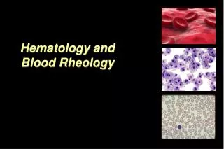
Hematology: the study ofBLOOD http://www.youtube.com/watch?v=YYqF78m5EW4 J. Hinson Human Anatomy and Physiology
I. Blood Overview • Connective tissue • Functions: • Transport substances b/w cells and the environment • Maintain a stable cellular environment • Heavier and more viscous than water
I. Blood Overview D. Composed of red blood cells, white blood cells, platelets, and plasma 1. 45% cells; 55% plasma 2. Differentiated from hemacytoblasts 3. Determined by CBC E. Normal blood volume: 4-6 L http://www.biosbcc.net/doohan/sample/images/blood%20cells/hematocrit.jpg
II. Red Blood Cells (Erythrocytes) • Shape allows for increase surface area for gas diffusion; lack nuclei • Hemoglobin (Hg): O2 carrying protein • Hematocrit (HCT): ratio of RBCs to total blood volume • RBCcount: # RBCs / mm3 - - - 4,200,000 – 6,200,000 RBCs/mm3 http://science.uwe.ac.uk/research/uploads/CRIB_blood_cells.jpg
Red Blood Cells (Erythrocytes)E. Hematopoeiesis: Red Blood Cell Production • Produced in red bone marrow • Negative Feedback Mechanism • Erythropoietin released from kidneys due to decrease in O2 • Red marrow increases RBC production • When RBC supply is sufficient to supply O2, erythropoietin release ceases. • RBC production is subsequently reduced.
II. Red Blood Cells (Erythrocytes) E. Hematopoiesis 3. Significantly affected by diet a. lack of B-complex vitamins, B12, and folate: pernicious anemia b. lack of iron: hypochromic anemia F. Life Span: ~120 days 1. Damaged cells are destroyed in liver and spleen by macrophages http://www.medicalook.com/systems_images/Platelets.jpg http://www.youtube.com/watch?v=_ZV5140OykE
III. White Blood Cells (Leukocytes) • Aid in immunity • WBC Count: 5,000-10,000 cells/mm3 1. Differential: WBC count (DIFF) http://diverge.hunter.cuny.edu/~weigang/Images/16-05_leukocytes_1.jpg
White Blood Cells (Leukocytes)C. Granulocytes v. Agranulocytes • Granulocytes: 2x the size of RBCs; contain cytoplasmic granules • Neutrophils • Eosinophils • Basophils • Agranulocytes: lack granules • Monocytes • Lymphocytes
White Blood Cells (Leukocytes)Neutrophils • Nucleus w/ 2-5 lobes; granules stain pink • 54-62% of WBC • Destroys relatively small particles by phagocytosis http://www.lab.anhb.uwa.edu.au/mb140/CorePages/Blood/blood.htm
White Blood Cells (Leukocytes)Eosinophil • Bilobed nucleus; coarse granules stain red • 1-3% of WBC • Kills parasites and helps control allergic reactions http://www.lab.anhb.uwa.edu.au/mb140/CorePages/Blood/blood.htm
White Blood Cells (Leukocytes)Basophil • Lobed nuclei (kidney shaped); irregular granules stain blue • <1% of WBC • Releases anticoagulant: heparin/histamine http://www.lab.anhb.uwa.edu.au/mb140/CorePages/Blood/blood.htm
White Blood Cells (Leukocytes)Monocyte • 2-3x larger than RBC; nuclei round or lobed • 3-9% of WBC • Destroys relatively large particles via phagocytosis http://www.lab.anhb.uwa.edu.au/mb140/CorePages/Blood/blood.htm
White Blood Cells (Leukocytes)Lymphocytes • Slightly larger than RBCs; large nuclei • 25-33% of WBC • Aid in immunity http://www.lab.anhb.uwa.edu.au/mb140/CorePages/Blood/Images/ly106le.jpg
IV. Platelets (thrombocytes) • Cytoplasmic fragments • Lack nuclei; < half size of RBCs • Platelet count: 130,000-360,000 platelets / mm3 • Function: repair breaks in damaged blood vessels and initiate clot formation http://diaglab.vet.cornell.edu/clinpath/modules/heme1/images/pltclmp.jpg
V. Plasma • Clear, straw-colored liquid suspension • 92% water • Functions: - transport nutrients, gases, and vitamins - regulation of fluid and electrolyte balance - maintenance of pH
V. Plasma D. Plasma proteins (produced in the liver) 1. albumins (60%): osmotic pressure and blood volume regulation 2. globulins (36%): transport lipids and aid in immunity 3. fibrinogen (4%): clotting E. Plasma nutrients: amino acids, simple sugars, and lipids F. Nitrogenous substances: amino acids, urea, uric acid G. Plasma electrolytes help maintain osmotic pressure and pH and include: Na+1, K+1, Ca+2, Mg+2, Cl-1, HCO3-1, PO4-3, and SO4-2
VI. Hemostasis: blood clotting • Smooth muscle contracts (vasoconstiction) to decrease blood loss. a. Platelets release serotonin to prolong. • Platelets adhere to collagen and stick to themselves forming a platelet plug.
VI. Hemostasis: blood clotting • Coagulation causes blood clot. Involves multiple clotting factors. • Prothrombin converted to thrombin. • Enzymatic thrombin converts fibrinogen to fibrin. • Meshwork traps platelets to form a blood clot. • Fibroblasts produce fibrous tissue which helps form a scab. http://www.youtube.com/watch?v=uBvpE5ia2LE&feature=channel
VI. Hemostasis: blood clotting • Some common coagulation disorders include: • Hemophilia B (Christmas Disease) • Von Willebrand’s disease B. Abnormal formation of a blood clot in a vessel is a thrombus. When a thrombus dislodges into blood flow, it is called an embolus. ** Often caused by fatty plaque/cholesterol deposits and may lead to death (ie stroke).
VII. Blood Typing • The process of identifying the antigens present on RBCs • Antigens are found on RBC; Antibodies are in the plasma. • ABO blood group: blood is typed based on the presence or absence of A or B antigens. ** Most common: O+ ** Least common: AB-
VII. Blood Typing C. Agglutinations: clumping of red cells following a transfusion reaction http://nobelprize.org/educational_games/medicine/landsteiner/index.html http://www.med-ed.virginia.edu/courses/path/innes/images/rcdjpegs/rcd%20agglutination%20x50b.jpeg
VII. Blood Typing D. Rh factor: presence or lack of Rh antigens 1. Rh negative can donate to Rh positive. However, the reverse can only occur once, as the body will develop Rh antibodies. ** First discovered in Rhesus monkeys, thus the “Rh”!
VII. Blood Typing • Erythroblastosis fetalis - condition in which agglutination of RBCs in an Rh+ fetus occurs during the second pregnancy of an Rh- mother * Rh antibodies developed during birth of the first fetus attack the second!