Download
1 / 44
440 likes | 461 Views
Learn about the Autonomic Nervous System (ANS) which regulates involuntary bodily functions without conscious control. Explore its components, functions, and the divisions – sympathetic and parasympathetic. Understand the pathways and effects of ANS activities in the body.

E N D
Introduction to ANS • Regulates activity of smooth muscle, cardiac muscle and glands • Operates without conscious control • Named autonomic because was thought to be AUTONOMUS (working without CNS)
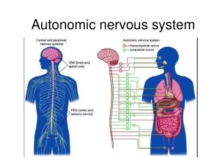
Autonomic Nervous System • But to operate it depends on continuous flow of sensory input from: • Visceral organs and • Blood vessels(not consciously perceived) into • INTEGRATING CENTERS IN THE CNS.
Autonomic Nervous System • Structurally then ANS includes: • Autonomic sensory neurons • Integrating centers in the CNS • Autonomic motor neurons to smooth muscle, cardiac muscle and glands Where in the CNS? Hypothalamus and brain stem
Comparing the ANS and Somatic NS • Structurally then SNS includes: • Somatic sensory neurons • Integrating centers in the CNS (Cortex) • Somatic motor neuron to skeletal muscles only
Comparing the ANS and Somatic NS • Somatic- the effect of a motor neuron is always excitation • Autonomic- the effect of a motor neuron is either excitatory or inhibitory
Autonomic Nervous System • Makes all routine adjustments in physiological systems. • The ANS pathway from the CNS to the effector always involves 2 neurons synapsing in an autonomic ganglion
ANS • Preganglionic (neuron #1) – cell body is in the CNS, axon extends to the ganglion outside the CNS • Postganglionic (neuron #2) – cell body is in the ganglion, axon extends to the visceral effector
Nerve Fibers of the ANS • Preganglionic (neuron #1) • Always myelinated • Neurotransmitter is always ACh • Postganglionic (neuron #2) • Always nonmyelinated • Neurotransmitter is Ach or norepinephrine
Subdivisions of the ANS • Sympathetic Division • Fight-or-flight • Parasympathetic Division • Rest-and-digest • These divisions are anatomically distinct
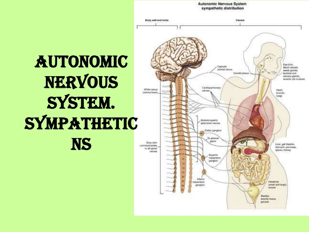
Sympathetic • Sympathetic division (thoracolumbar) • Cell bodies for all the neurons #1 reside in the thoracic and lumbar portions of the spinal cord. • T1 – L2
Sympathetic • Stimulates • heart beat • tissue metabolism, • increases alertness, • prepares the body to deal with emergencies • (“fight or flight” division)
Parasympathetic • Parasympathetic division (craniosacral) • Cell bodies reside in the brain stem (cranial nerves) or in the sacral portion of the spinal cord.
Parasympathetic • Slows the heart rate, • inhibits senses, • prepares the body for rest and relaxation; (“rest and digest” division).
Sympathetic Chain Ganglia • Synapses of neurons #1 and #2 are in a chain of ganglia that run alongside the spinal cord • Extends on both sides of the vertebral column • Carries preganglionic fibers and cell bodies of postganglionic neurons
Anatomy of the chain • Rami communicantes from the spinal nerves connect to the chain
Routes of Preganglionic Axons • Cell bodies of neurons #1 lie in the lateral gray horns of the spinal cord • The axons of neurons #1 leave the spinal cord via the ventral root • These axons pass to the spinal nerve • Axons leave the spinal nerve via the white branches (rami communicantes) • Connect with the sympathetic chain ganglia
Routes of Preganglionic Axons • There are 3 possible routes that sympathetic neurons may follow • Possibility #1: synapses within the ganglion at that level and • Second neuron leaves at that level via the gray ramus communicans, exits to the visceral effector
Routes of Preganglionic Axons • Possibility #2: neuron #1 goes up or down the chain and synapses at some other level. • Second neuron: leaves at that other level via the gray ramus communicantes, and exits to the visceral effector.
Routes of Preganglionic Axons • Possibility #3: neuron #1 does not synapse in the chain (exception!!) but exits and synapses in a collateral ganglion near a major blood vessel. • Neuron #2 travels from that ganglion to the visceral effector.
Where are the Collateral Ganglia ? • Location –Near a major blood vessel • Celiac ganglion • Innervates upper abdominal viscera • Superior mesenteric • Innervates middle abdominal viscera • Inferior mesenteric • Innervates lower abdominal & pelvic organs
The Adrenal Medulla • Yet another type of innervation: • Going to the adrenal medulla • No synapse in ganglia • No synapse in collateral ganglia • YES synapse in the adrenal medulla
Adrenal Medulla • Only preganglionic neurons are in this pathway • Neuron #1 stimulates the medulla, • The medulla releases norepinephrine and epinephrine (adrenaline) to blood
Effects of Sympathetic Stimulation • Widespread • The sympathetic chain allows one preganglionic fiber to synapse with many postganglionic neurons • Enhanced & prolonged by the adrenal medulla
Convergence • See heart
Neurotransmitters of Sympathetic Division • Preganglionic fibers release acetylcholine (Ach) Therefore they are called: • Cholinergic • Postganglionic fibers (most) release norepinephrine (NE) (=noradrenaline) • Adrenergic • Adrenal medulla releases norepinephrine and epinephrine (adrenalin)
Functions of the Sympathetic Division • Heart: increases rate • Lung bronchioles: dilates bronchioles • Salivary glands: produce viscous fluid • Stomach: decreases motility • Pupil: dilates • Sweat glands: produce secretions
Summary of Sympathetic Division • Cell bodies are found in the thoracic and lumbar portions of the spinal cord • Preganglionic fibers are short, connect to the sympathetic chain, and synapse with long postganglionic fibers • Preganglionic fibers produce ACh, postganglionic fibers produce NE or Ach • “Fight or flight” division
ANS either increases excitation or inhibits the activity • Ex. Sympathetic fibers increase heart rate, parasympathetic fibers decrease heart rate. • Homeostasis comes from the balance of the two.