Download
1 / 33
330 likes | 351 Views
Learn about achieving color consistency, issues of inconsistency in grayscale imaging, challenges of image presentation across devices, and solutions for maintaining image quality in medical imaging. This presentation covers topics such as color blending, hanging protocols, display calibration tools, and DICOM grayscale image transformations.

E N D
Consistent Presentation of Images Lawrence Tarbox, Ph.D. Washington University in St. Louis
Overview • Review of Grayscale Presentation State • Color Presentation States • Color Consistency • Presentation States applied to Color Images • Color Blending - CT-PET fusion • Hanging Protocols
Problems of Inconsistency • VOI chosen on one display device • Rendered on another with different display • Mass expected to be seen is no longer seen mass visible mass invisible Slide Provided by David Clunie, Quintiles Intelligent Imaging
0.5 1.0 • Not all display levelsare perceivable on alldevices 1.5 3.0 Problems of Inconsistency Slide Provided by David Clunie, Quintiles Intelligent Imaging
Digital Modality Laser Printer Problems of Inconsistency • Printed images don’t looklike displayed images Slide Provided by David Clunie, Quintiles Intelligent Imaging
Grayscale Standard Display Function Despite different change in absolute luminance 1000 100 10 1 0 200 400 600 800 1000 .1 .01 JND Index Same number of Just Noticeable Difference == Same perceived contrast Grayscale Standard Display Function Slide Provided by David Clunie, Quintiles Intelligent Imaging
Display Calibration Tools (Photometer) Slide Provided by Jerry Gaskill, Image Smiths Inc.
Monitor Characteristic Curve Monitor Characteristic Curve Luminance 100 10 0.1 Ambient Light 0.01 0 50 100 150 200 250 300 Digital Driving Level Slide Provided by David Clunie, Quintiles Intelligent Imaging
Mapping P-Values to Input of Characteristic Curve (DDL’s) 300 250 200 DDL 150 100 50 0 0 50 100 150 200 250 300 P-Values Perceptual linear device - LUT Slide Provided by David Clunie, Quintiles Intelligent Imaging
Grayscale Standard Display Function Despite different change in absolute luminance 1000 100 10 1 0 200 400 600 800 1000 .1 .01 JND Index Same number of Just Noticeable Difference == Same perceived contrast Grayscale Standard Display Function Slide Provided by David Clunie, Quintiles Intelligent Imaging
Laser Printer Digital Modality Workstation Workstation Distributed Image Consistency Identical perceived contrast
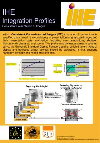
DICOM Grayscale Image Transformation Model Rescale Slope/Intercept or Modality LUT Window/Level or VOI LUT Presentation LUT Original Image Presentation LUT Transformation M o d a l i t y M a s k V O I L U T L U T Grayscale Transformations ( S u b t r a c t i o n ) T r a n s f o r m a t i o n T r a n s f o r m a t i o n S h u t t e r Display P-Values T r a n s f o r m a t i o n I m a g e D i s p . A r e a S p a t i a l Shutter, Annotation and Spatial Transformations T r a n s f o r m a t i o n A n n o t a t i o n A n n o t a t i o n
Spatial Transformations Entire Image Selected Flip Horizontal Scale To Fit Original Image Transformed Image
Spatial Transformations Part of Image Selected Flip Horizontal Scale To Fit Original Image Transformed Image
Transformation & Annotation Part of Image Selected Flip Horizontal Scale To Fit Mass behind heart Mass behind heart Original Image Transformed Image In this example, - text annotation is specified by image relative visible anchor point - the circle is a separate image relative graphic annotation
Printer Digital Modality Workstation Workstation Distributed Image Consistency Different perceived color
Printer Digital Modality Workstation Workstation Distributed Image Consistency Identical perceived color
Standard Color Space • GSDF filled a void • Color consistency already standardized • ICC - International Color Consortium • Graphics and pre-press industry • CIE Colorimetry • Profiles of input and output devices • COTS color management software handles conversion • Perceptual rendering intent
DICOM Grayscale Image Transformation Model Rescale Slope/Intercept or Modality LUT Window/Level or VOI LUT Presentation LUT Original Image Presentation LUT Transformation M o d a l i t y M a s k V O I L U T L U T Grayscale Transformations ( S u b t r a c t i o n ) T r a n s f o r m a t i o n T r a n s f o r m a t i o n S h u t t e r Display P-Values T r a n s f o r m a t i o n I m a g e D i s p . A r e a S p a t i a l Shutter, Annotation and Spatial Transformations T r a n s f o r m a t i o n A n n o t a t i o n A n n o t a t i o n
Blending for CT-PET selectunderlying selectsuperimposed
Blending for CT-PET selectunderlying [register] selectsuperimposed
Blending for CT-PET selectunderlying [register] selectsuperimposed resample
Blending for CT-PET selectunderlying [register] within slices selectsuperimposed resample
Blending for CT-PET selectunderlying [register] within slices selectsuperimposed resample [between slices]
Blending for CT-PET selectunderlying rescale andwindow [register] within slices selectsuperimposed resample [between slices]
Blending for CT-PET selectunderlying rescale andwindow [register] within slices selectsuperimposed pseudo-color resample [between slices]
Blending for CT-PET selectunderlying rescale andwindow blend [register] within slices selectsuperimposed pseudo-color resample [between slices]
Three New SOP Classes • Color Presentation State • Pseudo-Color Presentation State • Blending Presentation State • ICC Profile • Defines output of all color presentation states • Optionally present in all color images • PCS-Values (analogous to grayscale P-Values) • Profile Connection Space (CIELAB or CIEXYZ)
Hanging Protocols • “Default display protocols” • A set of instructions • How to layout a class of images for display • Order, orientation, windowing, processing • Not specific to a particular patient’s images • Hence a protocol, not a presentation state
L L L L Old Lateral New Frontal New Townes New Lateral F F F F New Study Old Study Hanging Protocols
Using a Hanging Protocol • Given a current exam (e.g. reading worklist) • Find potentially applicable protocols • Retrieve them from archive • Select one from those available • Select image +/- other studies to which it applies • Display selected images as instructed