Download
1 / 51
920 likes | 2.95k Views
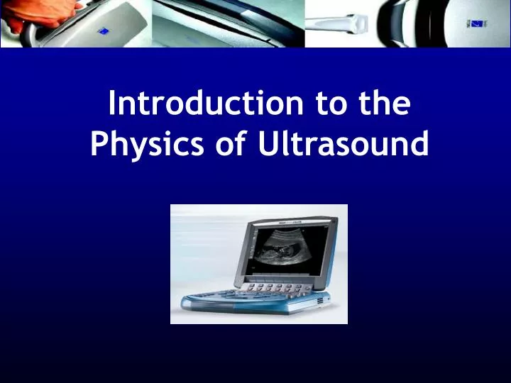
Introduction to the Physics of Ultrasound. ULTRASOUND PHYSICS. Format What is sound/ultrasound? How is ultrasound produced Transducers - properties Effect of Frequency Image Formation Interaction of ultrasound with tissue Acoustic couplants Image appearance . Sound?.

E N D
ULTRASOUND PHYSICS Format • What is sound/ultrasound? • How is ultrasound produced • Transducers - properties • Effect of Frequency • Image Formation • Interaction of ultrasound with tissue • Acoustic couplants • Image appearance
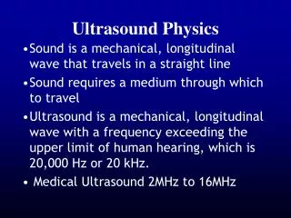
Sound? • Sound is a mechanical, longitudinal wave that travels in a straight line • Sound requires a medium through which to travel
Amplitude Basic Ultrasound Physics oscillations/sec = frequency - expressed in Hertz (Hz)
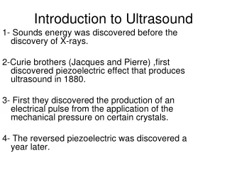
What is Ultrasound? • Ultrasound is a mechanical, longitudinal wave with a frequency exceeding the upper limit of human hearing, which is 20,000 Hz or 20 kHz. • Medical Ultrasound 2MHz to 16MHz
ULTRASOUND – How is it produced? Produced by passing an electrical current through a piezoelectrical crystal
Human Hair Single Crystal Microscopic view of scanhead
Piezoelectric material • AC applied to a piezoelectric crystal causes it to expand and contract – generating ultrasound, and vice versa • Naturally occurring - quartz • Synthetic - Lead zirconate titanate (PZT)
Ultrasound Production • Transducer contains piezoelectric elements/crystals which produce the ultrasound pulses (transmit 1% of the time) • These elements convert electrical energy into a mechanical ultrasound wave
The Returning Echo • Reflected echoes return to the scanhead where the piezoelectric elements convert the ultrasound wave back into an electrical signal • The electrical signal is then processed by the ultrasound system
Piezoelectric Crystals • The thicknessof the crystal determines the frequency of the scanhead Low Frequency 3 MHz High Frequency 10 MHz
Frequency vs. Resolution • The frequency also affects the QUALITY of the ultrasound image • The HIGHER the frequency, the BETTER the resolution • The LOWER the frequency, the LESS the resolution
Frequency vs. Resolution • A 12 MHz transducer has very good resolution, but cannot penetrate very deep into the body • A 3 MHz transducer can penetrate deep into the body, but the resolution is not as good as the 12 MHz
Broadband vs. Narrowband Amplitude Frequency
Broadband vs. Narrowband Nerve Visualisation: • 5-10 MHz • 6-13 MHz • By altering the transmit frequencies one transducer replaces several transducers • View a range of superficial to deep structures without changing transducers
Transducer Design Size, design and frequency depend upon the examination
Image Formation Electrical signal produces ‘dots’ on the screen • Brightness of the dots is proportional to the strength of the returning echoes • Location of the dots is determined by travel time. The velocity in tissue is assumed constant at 1540m/sec Distance = Velocity Time
Image Formation ‘B’ mode
Transducer Medium 1 Medium 2 Medium 3 Interactions of Ultrasound with Tissue • Acoustic impedance (AI) is dependent on the density of the material in which sound is propagated • - the greater the impedance the denser the material. • Reflections comes from the interface of different AI’s • greater of the AI = more signal reflected • works both ways (send and receive directions)
Interaction of Ultrasound with Tissue • Greater the AI, greater the returned signal • largest difference is solid-gas interface • we don’t like gas or air • we don’t like bone for the same reason • GEL!! • Sound is attenuated as it goes deeper into the body
Interactions of Ultrasound with Tissue • Reflection • Refraction • Transmission • Attenuation
transducer Interactions of Ultrasound with Tissue • Reflection • The ultrasound reflects off tissue and returns to the transducer, the amount of reflection depends on differences in acoustic impedance • The ultrasound image is formed from reflected echoes
Refraction reflective refraction Scattered echoes Incident Angle of incidence = angle of reflection
transducer Interactions of Ultrasound with Tissue Transmission • Some of the ultrasound waves continue deeper into the body • These waves will reflect from deeper tissue structures
Interactions of Ultrasound with Tissue Attenuation • Defined - the deeper the wave travels in the body, the weaker it becomes -3 processes: reflection, absorption, refraction • Air (lung)> bone > muscle > soft tissue >blood > water
Attenuation & Gain • Sound is attenuated by tissue • More tissue to penetrate = more attenuation of signal • Compensate by adjusting gain based on depth • near field / far field • AKA: TGC
Ultrasound Gain • Gain controls • receiver gain only • does NOT change power output • think: stereo volume • Increase gain = brighter • Decrease gain = darker
Balanced Gain • Gain settings are important to obtaining adequate images. bad far field bad near field balanced
Reflected Echo’s • Strong Reflections = White dots Diaphragm, tendons, bone ‘Hyperechoic’
Reflected Echo’s Weaker Reflections = Grey dots • Most solid organs, • thick fluid – ‘isoechoic’
Reflected Echo’s • No Reflections = Black dots • Fluid within a cyst, urine, blood ‘Hypoechoic’ or echofree
What determines how far ultrasound waves can travel? • The FREQUENCY of the transducer • The HIGHER the frequency, the LESS it can penetrate • The LOWER the frequency, the DEEPER it can penetrate • Attenuation is directly related to frequency
Ultrasound Beam Depth • Need to image at proper depth • Can’t control depth of beam • keeps going until attenuated • You can control the depth of displayed data
Ultrasound Beam Profile • Beam comes out as a slice • Beam Profile • Approx. 1 mm thick • Depth displayed – user controlled • Image produced is “2D” • tomographic slice • assumes no thickness • You control the aim 1mm
Goal of an Ultrasound System • The ultimate goal of any ultrasound system is to make like tissues look the same and unlike tissues look different
Accomplishing this goal depends upon... • Resolving capability of the system • axial/lateral resolution • spatial resolution • contrast resolution • temporal resolution • Processing Power • ability to capture, preserve and display the information
Types of Resolution • Axial Resolution • specifies how close together two objects can be along the axis of the beam, yet still be detected as two separate objects • frequency (wavelength) affects axial resolution – frequency resolution
Types of Resolution • Axial Resolution • specifies how close together two objects can be along the axis of the beam, yet still be detected as two separate objects • frequency (wavelength) affects axial resolution – frequency resolution
Types of Resolution • Lateral Resolution • the ability to resolve two adjacent objects that areperpendicular to the beam axis as separate objects • beamwidth affects lateral resolution
Types of Resolution • Spatial Resolution • also called DetailResolution • the combination of AXIAL and LATERAL resolution - how closely two reflectors can be to one another while they can be identified as different reflectors
Types of Resolution • Temporal Resolution • the ability to accurately locate the position of moving structures at particular instants in time • also known as frame rate
Types of Resolution • Contrast Resolution • the ability to resolve two adjacent objects of similar intensity/reflective properties as separate objects - dependant on the dynamic range
Ultrasound Applications Visualisation Tool: Nerves, soft tissue masses Vessels - assessment of position, size, patency Ultrasound Guided Procedures in real time – dynamic imaging; central venous access, nerve blocks
Imaging Know your anatomy – Skin, muscle, tendons, nerves and vessels Recognise normal appearances – compare sides!
Skin, subcutaneous tissue Epidermis Loose connective tissue and subcutaneous fat is hypoechoic Muscle interface Muscle fibres interface Bone
Transverse scan – Internal Jugular Vein and Common Carotid Artery
Summary • Imaging tool – Must have the knowledge to understand how the image is formed • Dynamic technique • Acquisition and interpretation dependant upon the skills of the operator.