Download
1 / 9
90 likes | 183 Views
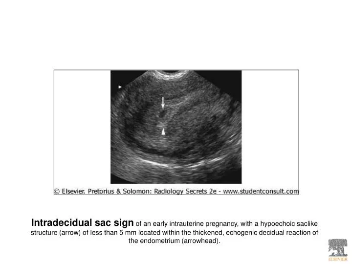
Intradecidual sac sign of an early intrauterine pregnancy, with a hypoechoic saclike structure (arrow) of less than 5 mm located within the thickened, echogenic decidual reaction of the endometrium (arrowhead).

E N D
Intradecidual sac sign of an early intrauterine pregnancy, with a hypoechoic saclike structure (arrow) of less than 5 mm located within the thickened, echogenic decidual reaction of the endometrium (arrowhead).
Normal early intrauterine gestational sac with separation of the hyperechoic decidual endometrial and gestational sac layers, creating the double decidual sac sign (arrows).
Normal 9-week intrauterine pregnancy shows a well-defined embryo (arrows) and yolk sac (arrowhead) with normal separation of the amnion (A) and chorionic (C) sacs.
Early failed intrauterine pregnancy with a calcified yolk sac (arrow) and embryonic remnant (arrowhead).
Color Doppler ultrasound of an incomplete abortion with increased vascularity, fluid, and echogenic debris (arrows) along the endometrium.
Grayscale (A) and Doppler (B) transvaginal ultrasound images of an ectopic (ECT) right adnexal pregnancy seen as a tubal ring separate from the right ovary (OV). There is also a small amount of adjacent particulate free fluid (FF).
Grayscale (A) and Doppler (B) transvaginal ultrasound images of an ectopic (ECT) right adnexal pregnancy seen as a tubal ring separate from the right ovary (OV). There is also a small amount of adjacent particulate free fluid (FF).
Pseudogestational sac (arrows) of an ectopic pregnancy with echogenic material consistent with hemorrhage filling the endometrial cavity.
Endometrial cavity filled with heterogeneous cystic and solid placental tissue (arrows), which is characteristic of a complete molar pregnancy.