Download
1 / 51
520 likes | 927 Views
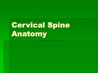
Cervical spine. Cervical Spine Anatomy. 1. 2. 3. 4. 5. 6. 7. Cervical Spine Anatomy. Vertebrae (7) Intervertebral discs (6) Pairs of exiting nerve roots (8) Cervical lordosis Occ-C7 averages 40° Most of the lordosis occurs at the C1-C2 segment. Cervical Spine Anatomy.

E N D
1 2 3 4 5 6 7 Cervical Spine Anatomy • Vertebrae (7) • Intervertebral discs (6) • Pairs of exiting nerve roots (8) • Cervical lordosis Occ-C7 averages 40° • Most of the lordosis occurs at the C1-C2 segment
Cervical Spine Anatomy • Approximately 50% of flexion-extension motion occurs at occiput-C1 • Approximately 50% of rotation occurs at C1-C2 • Lesser amounts of flexion-extension, rotation, and lateral bending occur segmentally between C2-C7
Cervical Spine Anatomy • Atypical vertebral • structure C1 (atlas) • Vertebral canal/foramen • Anterior arch • Anterior tubercle • Transverse process • Posterior arch • Transverse foramen • Lateral mass Inferior Superior
Cervical Spine Anatomy • Atypical cervical • vertebra C2 (axis) • Odontoid process or dens • Vertebral canal/foramen • Facet joints • Transverse process • Transverse foramen • Bifid spinous process • Lamina anterior view posterior view
Cervical Spine Anatomy • The odontoid process of the axis (C2) extends cranially to form the axis of rotation with atlas (C1)
Cervical Spine Anatomy • Ligaments • Anterior longitudinal ligament • Posterior longitudinal ligament • Ligamentum flavum • Intertransverse ligaments • Interspinous ligaments • Ligamentum nuchae
Cervical Spine Anatomy • Ligaments • Anterior longitudinal ligament • Posterior longitudinal ligament • Ligamentum flavum • Intertransverse ligaments • Interspinous ligaments • Ligamentum nuchae
Cervical Spine Anatomy • Ligaments • Anterior longitudinal ligament • Posterior longitudinal ligament • Ligamentum flavum • Intertransverse ligaments • Interspinous ligaments • Ligamentum nuchae
Cervical Spine Anatomy • Ligaments • Anterior longitudinal ligament • Posterior longitudinal ligament • Ligamentum flavum • Intertransverse ligaments • Interspinous ligaments • Ligamentum nuchae
Cervical Spine Anatomy • Ligaments • Anterior longitudinal ligament • Posterior longitudinal ligament • Ligamentum flavum • Intertransverse ligaments • Interspinous ligaments • Ligamentum nuchae
Cervical Spine Anatomy • Neural elements • 8 pair of cervical nerves • Exit the spinal canal superior to the vertebrae for which they are numbered • C1 nerves exit the canal between Occ & C1 • C2 nerves exit the canal between C1 & C2 • C8 nerves exit the canal between C7 & T1
Cause and discription Infantile (congenital) torticollis -common. The sternomastoid muscle on one side is fibrous and fails to elongate as the child grows; consequently, progressive deformity develops. The cause is unknown; the muscle may have suffered ischaemia from a distorted position in utero ( breech presentation and hip dysplasia ), or it may have been injured atbirth
Clinical feature - • -Hx-difficult labour or breech delivery • O/E –lump- there isneither deformity nor obvious limitation of movement- months the lump disapear ------Deformity does not become apparent until- • the child is 1–2 years old. The head is tilted to on one side, so that the ear approaches the shoulder; the • -sternomastoid on that side may feel tight and hard. • -There may also be asymmetrical development of the • face (plagiocephaly). become increasinglyobvious as the child grows
DDX lymphadenitis - -bony anomalies, -discitis, X_RAY
Treatment • -If the diagnosis is made during infancy-daily muscle stretching by the aparentsmay prevent the deformity. -If the condition persists beyondone year, operative correction is required -usually at its lower end but sometimes at the • upper end or at both ends) and the head is manipulated • into the neutral position. After operation, correction • must be maintained, with a temporary rigid • orthosis followed by stretching exercises.
Secondary torticollis infection (lymphadenitis, retropharyngeal abscess, tonsillitis, discitis, tuberculosis), trauma, juvenile rheumatoid arthritis, posterior fossa tumours, intraspinal tumours, dystonia (benign paroxysmal torticollis) or ocular dysfunction.
Cervical disc degeneration Cervical disc prolapse- CERVICAL SPONDYLOSIS- Cervical spondolytic myelopathy -ossification of posterior longitudinal ligament, cervical spinal canal stenosis--
Intervertebral Disc Hydrostatic, load bearing structure between the vertebral bodies Nucleus pulposus + annulus fibrosus No blood supply L4-5, largest avascular structure in the body
Nucleus Pulposus Type II collagen strand + hydrophilic proteoglycan Water content 70 ~ 90% Confine fluid within the annulus Convert load into tensile strain on the annular fibers and vertebral end-plate
Annulus Fibrosus Outer boundary of the disc More than 60 distinct, concentric layer of overlapping lamellae of type I collagen Helicoid pattern Resist tensile, torsional, and radial stress Attached to the cartilaginous and bony end-plate at the periphery of the vertebra
Vertebral End-Plate Cartilaginous and osseous component Nutritional support for the nucleus Passive diffusion
Facet Joint Synovial joint Rich innervation with sensory nerve fiber Same pathologic process as other large synovial joint Load share 18% of the lumbar spine
Vital Functions • -Restricted intervertebral joint motion • -Contribution to stability and-Preservation of anatomic relationship -Resistence to axial, rotational, and bending load
Nerve root • Medial & inferior to the pedicle at each level • More susceptiple for mechanical deformation
Introduction • Male predominance • 30 – 50 yrs • Smokers • Sudden flexion& Twisting
Intervertebral Disc Cellular and Biochemical Change Decrease proteoglycan content Water loss within the nucleus pulposus Decrease hydrostatic property Loss of disc height Uneven stress distribution on the annulus
Classification • A-Site;5-6,6-7 • B-Direction; posterolat • C-Amount ---Bulge --Herniation 1-Protrusion 2-extrusion 3- sequestration
Clinical picture Pressure on pll --------------- Dura Pressure on root Pressure on cord Mixed
EXAM • LOOK • FEEL • MOVE • NEUROLLOGICAL EXAM • SPECIAL TEST • -neck compression,distraction,valsava
Imaging • X-ray • MRI • CT scans with or without myelography -intolerant to MRI -Unsuitable for MRI • gadolinium-enhanced MRI This will help to delineate which part of the previous operation site is disc and which is epidural fibrosis (the latter enhancing).
DDX • Acute muscular&ST strain • Infection Tumor • Rotator cuff syndrome
Treatment • usually have a good prognosis • . In up to four-fifths of patients, symptoms will resolve spontaneously within a 12-week period. • However, if pain persists beyond this time there is a slow resolution of pain in the majority of patients.
By approximately 4 years there is no difference in the incidence of pain in patients treated non-operatively or surgically. • Surgical results will deteriorate after symptoms have been present for 1 year.
-REST-collar ANALGESICS&ANTIINFLAMATORY • ---Reduce-traction ----Remove -Rhablitate
Epidural steroid injection • If pain persist beyond 4 weeks • Maximum 3 injection per year
Indications for diskectomy • Strong indications for surgical intervention -Acute mylopathy or myloradiculopathy -Progressive Neurological deficit • Relative indications • Failure of conservative treatment-refractory • Significant motor deficit • Severe incapacitating pain - does not respond to any form of treatment