Download
1 / 31
310 likes | 426 Views
THE STUDY OF CELLS. 9 th Grade - Biology. INTRODUCTION. All Living organisms are made up of microscopic structural units called Cells. Diagram below shows the structure of generalized cell:-

E N D
THE STUDY OF CELLS 9th Grade - Biology
INTRODUCTION • All Living organisms are made up of microscopic structural units called Cells. • Diagram below shows the structure of generalized cell:- • We shall study about nucleus which is the most prominent cell organelle in more detail in the coming slides.
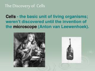
NUCLEUS • Nucleus isusuallyfound in the centre of the Cell. • Nucleus wasdiscovered by Robert Brown in 1831. • It issurrounded by a double layered membrane callednuclear membrane. This separates the substance in the nucleus from the cytoplasm. • The outer membrane iscontinuouswith the endoplasmicreticulum at certain places. • There are tiny pores callednuclear pore whichhelps in transportation of chemicalsusbtances in and out of the nucleus. • Nucleus contains a clear-jellylikeground substance callednucleoplasm or karyoplasm. It is made up of proteins and nucleicacids. • There are thread like structures in the nucleus whichform a structure calledchromatinthatappears as chromosomes duringcell division. Theytransferhereditarycharacteristicsfrom one generation to another. • Nucleolus, a darkspherical structure thatisfound in nucleus plays an important role in proteinsynthesis.
Chromosomes and the DNA • Nucleus is chiefly made up of proteins and nucleic acids. • There are 2 types of nucleic acids:- • Deoxyribo Nucleic acid / DNA • RiboNucleic acid / RNA • When the ultrastructure of chromosomes is observed, they appear as long spiral fibres. • They are made of proteins and DNA
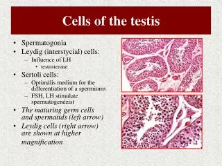
Structure of chromosomes • The proteins and DNA condense to formchromsomesduringcell division. • The number of chromosomes is for a particularspecies. For example, humancell has 46 chromosomes. DrosophilaMelanogaster, a fruit fly has 8 chrmosomes. • The chromosomes willbe in pairs always and in each pair twoidentical chromosomes are present. Hencethey are calledhomologous chromosomes. • In a pair of homologous chromosomes, each chromosome will have two parallel strands called chromatids. These 2 chromatids are held together at a point called centromere.
Structure of Chromosomes • The structure of a chromosome reveals that it is made up of proteins and a long tightly coiled DNA molecule. • The DNA molecule consists of thousands of hereditary units called genes. • There are genes to control each and every trait of an individual. • The genes control the structure and function of cells in all organisms from amoeba to man. • Some specific genes are responsible for colour of the eyes like blue eyes, green eyes, curly hair, colour of the skin, height and many such traits in man.
Deoxyribo nucleic acid - DNA • American scientist James Watson and an English scientist Francis Crick explained the struture of DNA molecule in 1953. • They were awarded the nobel prize in 1962 for their significant contributions. • They proposed a model called the Watson-Crick model or the double-helix model to describe the structure of DNA. • The structure of DNA resembles a twisted ladder which is known as the double-helix. • The 2 strands of DNA are built of large no of small units called nucleotides. • A single nucleotide consists of a deoxyribose sugar, a phosphate unit and a nitrogen base. • The deoxyribose sugar is a pentose sugar. • Each strand of the ladder is made up of deoxyribose sugar and phosphate units arranged alternately. • The nitrogen bases connect the opposite strands.
Deoxyribo nucleic acid - DNA • There are four types of nitrogen bases:- • adenine • Thymine • guanine • cytosine. They are represented as A,T,G and C respectively. • Adenine and guanine are called purines. • Cytosine and thymine are called pyramidines. • A purine on one chain always pairs with a pyramidine on the other chain, • Adenine pairs only with thymine and guanine pairs only with cytosine. Hence, the two strands of DNA are not identical but they are complementary to one another.
SIGNIFICANCE OF DNA • A living cell has to carry on a number of metabolic activities • It requires enormous information and instructions. All this information is coded in the DNA molecule. • This genetic information is passed on from generation to generation. To put it briefly, DNA controls all the activities of the cell. • The unique feature of DNA is its property of duplicating itself during cell division. This property is known as replication. • This special property of DNA is responsible for equal distribution of genetic material from the parent cell to the two daughter cells during cell division. • Please note that Adenine and thymine are heid together by two hydrogen bonds. Guanine and Cytosine are held together by three hydrogen bonds. • Any sudden change that occurs in the structure of DNA is called mutation
SIGNIFICANCE OF DNA • DNA also plays an important role in the synthesis of proteins. • It undergoes mutations and causes variations in organisms which lead to evolution of species. Thus DNA is mainly responsible for heredity and variation. • It is the blueprint for all life processes that take place in the cell in all living organisms. • To pass on DNA from one cell to another, a process called cell division occurs. • We have observed that a torn skin in a wound is replaced by a new skin within a short period. We have also observed a small baby growing into a youth. Unicellular organisms increase In their number. • All these activities of the living organisms involve the basic process in which a cell, growing for smetime, reaches maturity and then divides and produces daughter cells. This process is known as cell division.
CELL DIVISION • Cell division is a process by which cells reproduce their own kind. • In multicellular organisms growth, reproduction and repair take place through cell division. • There are two types of cell division. • Mitosis • Meiosis
MITOSIS • Mitosis is the most common type of cell division observed in higher plants and animals. Since it occurs in vegetative cells, it is called somatic cell division. • The changes that take place before and during Mitotic cell division are divided into two major phases. • Nuclear division (karyokinesis) • Cytoplasmic division (cytokinesis) • Initially the nucleus of the cell divides into two. This phase is called nuclear division or karyokinesis. • It is followed by the division of the cytoplasm. This phase is called cytokinesis. • A number of activities like storage of food and sythesis of materials take place inside the cell. just before the cell division. This phase is called Interphase. • During this phase, DNA found in every chromosome replicates to ensure equal distribution of the genetic material to the future daughter cells.
KARYOKINESIS • Karyokinesis can be split into 4 phases:- • Prophase 2. Metaphase 3. Anaphase 4. Telophase • Prophase • It is the longest phase. • The chromatin network becomes reorganized into chromosomes. • The DNA molecule coils tightly to form chromosomes. Here, it is to be noted that DNA changes physically but its chemical constitution remains the same. • Each chromosome appears to be made up of two strands. These parallel strands are called chromatids. • The two chromatids in a chromosome are held together at a point called centromere. • The centrioles that are normally found above the nucleus, move to the opposite poles of the cell. They develop cytoplasmic fibres from microtubules and appear as stars. They are now called asters. Asters are not formed in plant cells. • Long cytoplasmic fibres appear between the two asters. They are called spindle fibresbecause they look like a spindle. • The nucleolus disappears. • The nuclear membrane distintegrates. • The chromosomes are set free in the cytoplasm.
KARYOKINESIS • Metaphase • It is the phase of the shortest duration. • The chromosomes move towards the centre of thercell. • They arrange themselves in the equatorial plane. • This arrangement is also called metaphase plate. • The chromosomes are attached to the spindle fibre by their centromeres. • The lining up of the chromosomes mark the plane along which the cell divides
KARYOKINESIS • Anaphase • During this phase, the spindle fibres contract and the centromeres holding the chromatids of each chromosome, split into two. • Each chromatid gets one centromere. • The two chromatids separate and begin to move away to the opposite poles. • The spindle fibres attached to the centromeres pull the chromosames to their respective poles. • In this phase, the two sets of chromosomes separate. • A new set of interzonalfibresdevelop between the two poles.
KARYOKINESIS • Telophase • It is the final phase of karyokinesis. • In this phase the chromosomes reach the poles. • They uncoil and become thread like structures forming chromatin network. • A nuclear membrane appears around this network forming a nucleus at each pole. • A nucleolus appears. • The spindle fibres disappear. • Thus, two exactly similar nuclei are formed at the end of karyokinesis.
CYTOKINESIS • It is the division of the cytoplasm . • In the animal cell, a cleavage appears around the cell. It deepens as if a string is tightened around the middle of the cell. The groove formed is called cleavage furrow (2.6). • In the plant cell, a cell plate appears as a faint line at the equator. It gradually develops into a cell wall. • Thus, at the end of mitosis, two identical daughter cells containing the same number of chromosomes are formed. Hence, mitosis is called equational division. • The parent cell and each of the two daughter cells have the same number of chromosomes. This number is called diploid number which is represented as 2n (n= number of chromosomes). • Every human cell contains 23 pairs of chromosomes out of which 22 pairs are somatic chromosomes and one pair are sex chromosomes. We now know that, mitosis takes care of equal distribution of chromosomes to the daughter cells. The diploid number of chromosomes is maintained.
SIGNIFICANCE OF MITOSIS • Genetic stability : Since DNA replicates during mitosis forming exact copies, the genetic information received by the daughter cells will be the same. The daughter cells receive the same number of chromosomes which are genetically identical. Hence, genetic stability is maintained. • Growth : Unicellular organisms increase their number through mitotic divisions. In multicellular organisms growth and development of the body take place as a result of mitosis and increase in number of cells. • Cell Replacement : Cells will be constantly dying and disintegrating in the body. They have to be replaced. Replacement of cells and tissues in the body involves mitosis. • Healing : Mitosis is essential for healing of the wounds and repairing the worn and torn parts. • Regeneration : Animals like star fish can grow whole parts of the body if they are cut through mitosis. CANCER • When cells lose control over division, they result in tumors. If the cells in tumor can separate and divide again, they become malignant tumors that can cause cancer.
MEIOSIS • During sexual reproduction, male and female reproductive cells unite to form a zygote. • Zygote contains diploid number of chromosomes. • It is observed that the reproductive cells undergo a special type of cell division before the formation of zygote, during sexual reproduction. This cell division is called meiosis. • We know that somatic cells contain diploid number of (2n) of chromosomes. • However, the reproductive cells (germ cells) undergo meiosis in order to reduce the diploid number of chromosomes to half the number known as haploid number. It is represented as 'n'. • Hence, meiosis is also known as reductional cell division. • The cells with haploid number of chromosomes are called gametes. • When the male and female gametes unite to form the zygote during fertilization, 2n, the diploid number, is restored. Thus, the number of chromosomes for a particular species remains constant.
STAGES OF MEIOSIS • Meiosis involves two successive cell divisions. The two divisions are called • Meiosis I • Meiosis II • Meiosis I is reductional division and Meiosis II is equational division. • Meiosis I also involves karyokinesis (nuclear division) and cytokinesis (cytoplasmic division) just as in mitosis. DNA replicates during the interphase. • Meiosis I is immediately followed by Meiosis II MEIOSIS I • The karyokinesis in meiosis I is divided into four phases:- • prophase I • metaphase I • anaphase I • telophase I
MEIOSIS – Prophase I • In this phase, the chromatin material reorganizes into chromosomes. The nucleolus disappears. The nuclear membrane disintegrates. • Spindle apparatus is formed. • The homologous chromosomes pair with each other. The pairing of homologous chromosomes is called synapsis. • Each chromosome vertically splits forming two Chromatids. • Each pair of homologous chromosomes consists of four chromatids. This stage is known as tetrad. • In these 2 pairs of chromosomes, one pair comes from mother and the other from father. • Usually, the chromosomes will be of the same length in each pair andthe genes are arranged in the same order. Their centromeres will be at the same point. • The paired chromosomes are joined at one or more points along their length. These points are called chiasmata. • The chromosomes exchange genes at these points forming gene combinations. This process is called crossing over. • Following crossing over, the chromosomes repel each other and each pair assumes a shape depending on the number of chiasmata.
MEIOSIS – Metaphase I • The chromosomes move towards the equatorial plane. • They are attached to the spindle by their centromeres. • The centromeres of the homologous chromosomes lie on either side of the equator.
MEIOSIS – Anaphase I • In this. phase, one set of homologous chromosomes move towards one pole and another set towards the opposite pole. • The centromeres do not divide. • The haploid chromosomes are slightly different in their gene structure when compared to those of parent cells because of crossing over. • This difference accounts for the variations seen between parent organisms and their offspring.
MEIOSIS – Telophase I • The haploid chromosomes reach the opposite poles. • The chromatids are not separated. • A second meiotic division is necessary to separate the chromatids. • The chromosomes at each pole unwind and become thread like structures. • Nuclear membrane appears and two nuclei are formed. • Meiosis II will occur simultaneously in both the haploid cells
MEIOSIS – Prophase II • Meiosis II: Even in meiosis II, karyokinesis and cytokinesis occur. Karyokinesis is divided into four phases namely:- • prophase II • metaphase II • anaphase II • telophaseII. • Prophase II: • The changes that take place during this phase are similar to those of mitotic prophase. • However, a single homologue of each chromosome is present in each cell. • Each cell contains two sister chromatids joined by centromeres. • The centrioles move towards opposite poles. Spindle fibres appear. • The chromosomes are arranged at right angles to the spindle of meiosis I.
MEIOSIS – Metaphase II • The chromosomes arrange along the equator of the cell. • The spindle fibresare attached to the centromeres.
MEIOSIS – Anaphase II • Anaphase II is different from anaphase I. • The centromere divides into two and the two chromatids separate. • The sister chromatids of each chromosome move towards opposite poles along the spindle fibres.
MEIOSIS – Telophase II • The movement of chromosomes complete forming a nuclei. • The nuclear membrane and nucleolus appear. • The spindle fibres disappear. • Thus, a single diploid cell produces four haploid cells. • In plants the haploid cells are called spores. In animals the haploid cells are called gametes.
SIGNIFICANCE OF MEIOSIS • The diploid number of chromosomes (2n) is reduced to haploid number (n) in reproductive cells. • Meiosis brings about genetic variations due to exchange of genes during crossing over between the two parental (male and female) chromosomes. • It helps in maintaining a constant diploid number of chromosomes for a species.