Download
1 / 27
280 likes | 606 Views
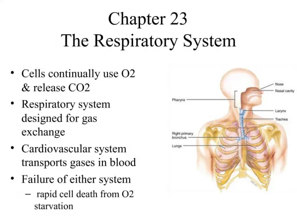
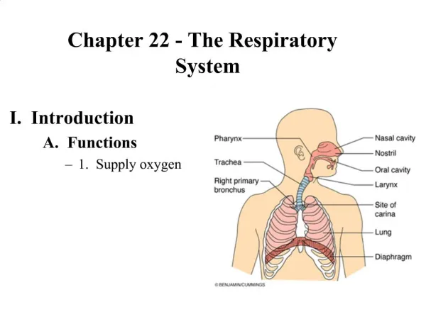
Chapter 21 – The Respiratory System . Nose . External nose is composed of 2 nasal bones (root/bridge) and hyaline cartilage Internal nasal cavity is divided into left and right sides by nasal septum Superiorly – perpendicular plate of the ethmoid bone; inferiorly – vomer. Nose cont.

E N D
Nose • External nose is composed of 2 nasal bones (root/bridge) and hyaline cartilage • Internal nasal cavity is divided into left and right sides by nasal septum • Superiorly – perpendicular plate of the ethmoid bone; inferiorly – vomer
Nose cont • Lined with mucous membrane • Olfactory – chemoreceptors for sense of smell • Respiratory • Pseudostratified columnar epithelium • Ciliated to trap debris • Mucous glands/goblet cells • Serous glands – lysozyme
Nose cont • Lateral walls have projections – conchae • Superior and middle conchae are part of the ethmoid bone; inferior conchae are paired bones • Increases air turbulence • Large particles get trapped in mucus • Warms and moistens air • Better gas diffusion
Paranasal sinuses • Frontal, maxillary, ethmoid, and sphenoid bones • Warms and moistens air • Lightens skull bones • Resonance chambers
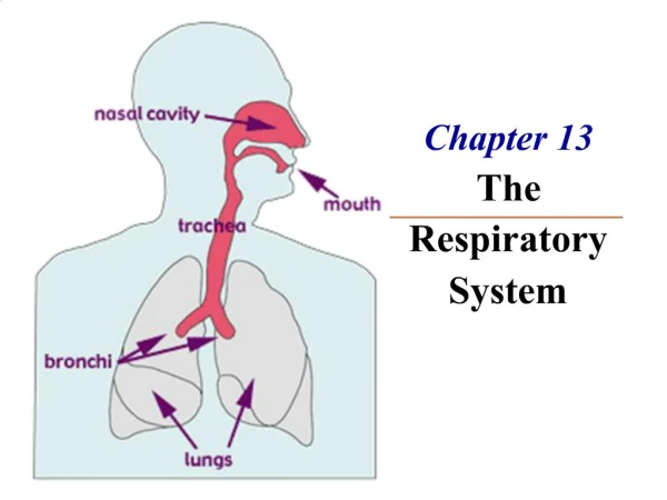
Pharynx • Connects nasal cavity with inferior respiratory structures • Common to both digestive and respiratory systems • From superior → inferior • Nasopharynx • Oropharynx • Laryngopharynx
Pharynx cont • Nasopharynx • Passageway for air only (not shared with digestive system) • Contains singular pharyngeal tonsil (“adenoid”) • Lymphoid material • Swelling can cause restriction of air flow • Contains opening to auditory/pharyngotympanic tube
Pharynx cont • Oropharynx • For both air and food/fluid • Contains paired palantine and paired lingual tonsils • Tissue changes to stratified squamous • Offers more protection • Laryngopharynx • For both air and food/fluid • Splits into anterior larynx (respiratory system) and posterior esophagus (digestive system)
Larynx • Supported by cartilage • Epiglottis • Moves inferiorly during swallowing to cover opening of larynx – food or fluid enters esophagus • Thyroid cartilage • “Adam’s apple” • Cricoid cartilage – complete ring • Arytenoid cartilage – attaches to vocal cords • Corniculate – “little horns”
Larynx cont • Voicebox • True vocal cords – inferior • False vocal cords/vestibular cords – superior • Aid in closing of the glottis – opening between true vocal cords
Trachea • “windpipe” • Ciliated pseudostratified • Supported by C-rings of cartilage • Incomplete posteriorly; allows esophagus to expand anteriorly when swallowing • Carina – projection of cartilage just before split into primary bronchi • Initates coughing when touched
Bronchi • Trachea branches into left and right primary bronchi • Each enters a lung at hilum (depression) • Right primary is wider, shorter, and more vertical • Food can get lodged there more easily • Each primary bronchus branches into secondary/lobar bronchi • 3 on right; 2 on left
Bronchi cont • Bronchi branch repeatedly • Transition of cartilage rings to cartilage plates • Lead to smaller bronchioles • Terminal bronchi → terminal bronchioles → respiratory bronchioles • Respiratory bronchioles have scattered alveoli • Alveolar sacs resemble clusters of grapes
Alveoli • Type I cells • Simple squamous epithelium • Site of gas exchange • Surrounded by capillary beds • Type II cells • Cuboidal cells • Secrete surfactant • Keep alveoli from collapsing • Alveolar pores connect adjacent alveoli so gas pressure is equalized • Macrophages on interior of alveoli
Zones • Conducting zone • Nasal cavity to terminal bronchioles • Passageway for gases only • Respiratory zone • Respiratory bronchioles to alveoli • Actual site of gas exchange
Lungs • Apex – most superior portion; base – most inferior (lies on top of diaphragm) • Medial surface has a depression – hilum • Entrance of primary bronchi, passage for blood vessels • Left lung • 2 lobes (superior and inferior) separated by oblique fissure • Cardiac notch • Depression to accommodate heart • Right lung • 3 lobes (superior, middle, and inferior) • Oblique and horizontal/transverse fissures
Lung blood supply • Pulmonary circulation • Pulmonary arteries – oxygen-poor blood to alveolar capillaries • Pulmonary veins – oxygen-rich blood back to heart • Systemic circulation • Bronchial arteries – branch from aorta to supply lung tissue (except alveoli) • Oxygen-poor blood from lung tissue enters small bronchial veins
Pleural layers • Parietal – more superficial • Lines thoracic wall and superior portion of diaphragm • Visceral – deep • Right on top of lung surface; dips into fissures • In between layers is pleural cavity • Pleural fluid for lubrication
Respiratory system function • Supplies blood with oxygen and removes waste carbon dioxide • 4 stages • Pulmonary ventilation • “breathing” • External respiration • Gas exchange at capillaries covering alveoli • Oxygen loading; carbon dioxide unloading • Transport of respiratory gases • By cardiovascular system • Internal respiration • Gas exchange at systemic capillaries • Oxygen unloading; carbon dioxide loading
Mechanics of Breathing • Pressure relationship • Atmospheric pressure – 760mm Hg • Intrapulmonary pressure – inside alveoli • Intrapleural pressure – inside pleural cavity • Differences between intrapulmonary and intrapleural pressures keep airways open • Elasticity of lungs – recoil into small shape; surface tension of surfactant keeps alveoli small • Opposition to this is the expansion of the thoracic cavity, which increases volume
Pulmonary ventilation • Boyle’s law • At constant temperature, gas pressure is inversely proportional to volume • Change in lung volume will affect lung pressure • Pressure changes leads to gas movement (high pressure area to low pressure area
Pulmonary ventilation cont • Inspiration • Diaphragm contracts from upward dome to flattened shape • Increases height of lungs • Contraction of intercostal muscles causes ribcage to swing outward • Increases depth of lungs • Lung volume increases, which decreases pressure in lungs • Pressure is now less than atmospheric pressure • Gas rushes into lungs • Expiration • Relaxation of diaphragm and intercostals allows lungs to recoil and assume smaller volume • Increase in pressure causes gas to rush out
Gas exchange • Basic gas properties • Dalton’s law of partial pressure • In a mixture of gases, each gas exerts pressure based on its relative % • Partial pressure • Henry’s law • When gas comes in contact with a liquid, the gas will dissolve into the liquid • If partial pressure of the gas is greater in liquid, gas will resume gaseous form and exit from liquid
Gas exchange cont • Carbon dioxide is most soluble in water; oxygen 1/20 of carbon dioxide • External respiration • Pressure of oxygen in alveolar capillaries is low compared to in alveoli • CO2 + H2O ↔ H2CO3 (carbonic acid) ↔ HCO3-(bicarbonate ion) + H+ (hydrogen ion) • Carbon dioxide is in 3 forms in blood • Bicarbonate ions • Carbaminohemoglobin • Dissolved in plasma • Partial pressure of carbon dioxide is higher in alveolar capillaries compared to alveoli • Internal respiration • Opposite movement of external respiration
Control of respiration • Brainstem • Detects rising CO2 levels in blood –causes drop in pH (too acidic) • Medulla oblongata • Controls rate/rhythm of breathing by contraction of diaphragm and external intercostals • Pons • Smooth out signal sent by medulla • Chemoreceptors • In aortic arch and carotid bodies (branch of internal and external) • Detect both rising CO2 levels and dropping O2 levels • Sends appropriate signal to brainstem to increase rate of respiration