Download
1 / 179
1.95k likes | 2.51k Views
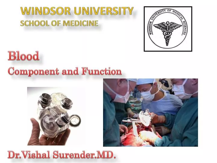
WINDSOR UNIVERSITY SCHOOL OF MEDICINE . Blood Component and Function Dr.Vishal Surender.MD. OBJECTIVES: BLOOD COMPOSITION. Describe the components of blood (cells, ions, proteins, platelets) giving their normal values. Describe the physico -chemical properties of the plasma .

E N D
WINDSOR UNIVERSITYSCHOOL OF MEDICINE Blood Component and Function Dr.Vishal Surender.MD.
OBJECTIVES: BLOOD COMPOSITION • Describe the components of blood (cells, ions, proteins, platelets) giving their normal values. Describe the physico-chemical properties of the plasma. • Define the term hematocrit, state its normal values. Explain the importance of maintaining normal hematocrit level. List the factors that affect the hematocrit value. Understand how dehydration, excessive water intake, RBC count and size affect Htc. Be able to calculate plasma volume for given Htc and total blood volume for given Htc and plasma volume. • Describe the functions of plasma electrolytes. • List the main fractions of plasma proteins and describe their properties and functions. Describe the reasons and consequences of hypoproteinemia. Understand why liver diseases with decreased synthesis of albumins cause edema formation. What is the meaning of the term ‘non-protein nitrogen’?
THE COMPONENTS OF BLOOD Blood is an opaque, red liquid consisting of several types of cells suspended in a complex, amber fluid known as plasma. When blood is allowed to clot or coagulate, the suspending medium is referred to as serum.
BLOOD FUNCTIONS • Transportation • Dissolved or chemically bound matter (O2, CO2, nutrients, metabolites) • Heat for heating and cooling (maintenance of the body temperature) • Protection/Immunity • Defense against foreign agents (specific and non-specific immunity) • Hemostasis • Prevention of hemorrhage - hemostasis • Regulation/Hemostasis • Transmission of signals (hormones) • Maintenance of the homeostasis • Concentration of dissolved substances • Osmotic and oncotic pressure • Acid-base balance - buffering the body fluids (maintenance of pH)
BLOOD VOLUME AND COMPOSITION Blood volume Males: 5000 – 6000 mL Females: 4000 – 5000 mL 6 - 8% of the total body mass 20% of the ECF. Composition • Blood plasma - non-cellular portion of the blood - 55% 2. Formed elements (45 %): a. Red blood cells, RBC (erythrocytes) -99% of formd. Elmts. b. Platelets (thrombocytes) c. White blood cells, WBC (leukocytes)
Blood Composition Plasma Blood HEMATOCRIT (Htc) Note that hematocrit is also known as Packed Cell Volume (PCV) or Erythrocyte Volume Fraction (EVF)
Composition of Blood Plasma Amino acids Water Albumin ions Globulin Proteins Fibrinogen Organic molecules Glucose such as is composed of PLASMA Lipids Trace elements and vitamins Nitrogenous wastes CO2 Gases such as O2 Figure 16-1 (1 of 2)
BLOOD PLASMA Physico-chemical properties • Temperature: 38oC • pH: 7.35 (venous) – 7.45 (arterial) • Relative whole blood viscosity - the internal friction of the blood (viscosity of water is 1.0): 3.5 – 5.4 • Relative plasma viscosity: 1.9 – 2.6 • Erythrocyte sedimentation rate- 2 to 8 mm/hr. Composition • Water: 90-92% • Functions • Solvent & suspending medium for blood components • Absorbs, transports and release heat • Electrolytes(Na+, K+, Ca2+, Fe3+, Mg2+, Cl-, HCO3-, HPO42-, SO42-etc.) dissolved in water: 1% • Functions • Create & maintain plasma osmotic pressure - 290 mOsm/L • Essential role in cell functioning (i.e., electrical properties of the blood cells) • Maintenance of the acid-base homeostasis*- • is a steady state that provides an optimal internal environment for cell function
BLOOD PLASMA: COMPOSITION (cont.) • Organic substances (plasma proteins, nutrients, metabolites and waste products, regulatory substances, etc.) – 7-9% • Plasma proteins – proteins confined to the blood • Nutrients - products of digestion (AA, glucose, FA, glycerol, vitamins, minerals) • Are transported by the blood for distribution in the body • Waste products – breakdown products of protein metabolism (i.e., urea, uric acid, creatine, creatinine, bilirubin and ammonia) • Are transported by the blood for excretion from the body • Regulatory substances (i.e., hormones, enzymes, vitamins) • Dissolvedgases: O2, CO2, N2
PLASMA PROTEINS The most charged proteins • 7-9% of the plasma, 65-90 g/L • Are synthesized by the liver (with the exception of γ-globulins) • Fractions • Albumins – plasma concentration – 45 g/L • Globulins (α, β, γ) – 27 g/L • Fibrinogen – 3 g/L • Albumins have the smallest molecular mass whereas fibrinogen is the largest The least negatively-charged serum proteins
GENERAL FUNCTIONS OF PLASMA PROTEINS • Create colloid osmotic pressure (25 mmHg) →retaining water within the capillaries • Binding and transport of hormones, enzymes, lipids, vitamins, metals, bilirubin, drugs, etc. • Contribution to the blood viscosity • Buffer properties – capability of accepting both H+ and base ions • Protection of body against microorganisms and toxic substances • Mediate blood coagulation • Precursors of some hormones (angiotensinogen, erythropoietin) • Protein reserve – source of AA for tissues in case of starvation
PROPERTIES & FUNCTIONS OF VARIOUS FRACTIONS OF PLASMA PROTEINS • Albumins • 60% of the total plasma protein • High concentration and small size → 80% of the total colloid osmotic P of the plasma • 1 globulins (glycoproteins) • Transport of glucose and lipids • Include anti-protease • 2 globulins • Carriers for different substances(high affinity, low binding capacity) • Ceruloplasmin – copper • Thyroxin-binding protein • Transcobalamin – Vit B12 • Bilirubin binding globulin • Transcortin – cortisol, etc.
PROPERTIES & FUNCTIONS OF VARIOUS FRACTIONS OF PLASMA PROTEINS (cont.) • -globulins • Carriers for lipids (lipoproteins), polysaccharides and metals (i.e., transferrin – iron and cupper) • -globulins • Are immunoglobulins (antibodies) • Quantity and composition fluctuate • ↑ in almost all diseases (inflammation and infections) • Fibrinogen • A dissolved precursor of fibrin – blood clotting • Serum – plasma without fibrinogen (and clotting factors)
HYPOPROTEINEMIA • ↓ blood level of proteins • Results from • Malnutrition • Liver diseases (depression of protein synthesis) • Intestinal disease (malabsorption) • Kidney diseases (lost of albumins in urine) • Results in • ↓ plasma oncotic pressure(especially due to ↓ albumin concentration) and edema formation • Depression of specific functions (i.e., ↓ in globulins – ↓ resistance to infections, ↓ in fibrinogen – defective blood clotting)
NON-PROTEIN NITROGEN OF THE PLASMA • Refers to nitrogen-containing substances other than proteins and AA (urea, uric acid, creatinine) • ↑ in deranged kidney function
HEMATOCRIT (Htc)-Important Diagnostic Measurement • Is the fraction of the blood volume made up of the formed elements (mainly RBC) • Is determined by the centrifuging heparinised/anticoagulatedblood in a standard calibrated tube of a small diameter • When blood is allowed to clot or coagulate, the suspending • medium is referred to as serum
(Htc)-Important Diagnostic Measurement Plasma (55% of whole blood) Buffy coat: Leucocytes and Platelets <1% of whole blood Formed Elements Erythrocytes (45% of whole blood)
HEMATOCRIT Determination of hematocrit values is a simple and important screening diagnostic procedure in the evaluation of hematological disease • Values • Males: 40 – 54 vol% (mean – 47%; 0.47) • Females: 38 – 46 vol% (mean – 42%, 0.42) • ↑ in persons leaving at high altitudes, in dehydrated state, polycythemia, etc. • ↓ in anemia, leukemia, bone marrow failure • Importance • Determines blood viscosity • ↑ Htc → ↑ resistance to blood flow, load on the heart & BP The contribution of the WBC to hematocrit is only 0.08%. WBCs are lighter than the RBCs, they form a thin whitish layer between the sedimented RBCs and the plasma.
HEMATOCRIT Tube A Tube B Tube C Normal Anemia Polycythemia
CONTENT • RED BLOOD CELLS (RBC) COUNT, FUNCTIONS, STRUCTURE • HEMOGLOBIN (Hb): CHEMISTRY, REACTIONS, FUNCTIONS, CONCENTRATION • ERYTHROPOIESIS, CONTROL OF ERYTHROPOIESIS • DESTRUCTION OF RBC, METABOLISM OF Hb AND IRON. HEMOSIDEROSIS • JAUNDICE • ERYTHROCYTE SEDIMENTATION RATE • TYPES OF ANEMIA, SICKLE CELL DISEASE • POLYCYTHEMIA
OBJECTIVES • Describe the functional consequence of the lack of a nucleus, ribosomes, and mitochondria for a) protein synthesis and b) energy production within the red blood cell. • Relate the three red blood cell concentration estimates, red blood cell count, hematocrit, and hemoglobin concentration. • Know the importance of MCV and be able to calculate the mean corpuscular volume. • Describe the structure of hemoglobin (Hb). Describe the differences between the major normal types of Hb (adult A and A2, glucosilated, fetal). Predict the changes in Hb types present in blood when synthesis of beta chains of globin is deficient. Describe the abnormal types of Hb (Hb S, thalassemias). Describe the normal and abnormal Hb reactions (oxyHb, MetHb, carboxyHb). Calculatethe mean corpuscular Hb concentration and the mean corpuscular Hb. • Identify the site of erythropoietin production, the adequate stimulus for erythropoietin release, and the target tissue for erythropoietin action. Describe the role of vitamin B12 & folic acid, and various hormones in regulation of RBC formation. Describe the dietary requirements for RBC production. Relate the rate of red blood cell production and the percentage of immature reticulocytes in the blood. • Describe the metabolism of iron in the body. • Describe the metabolism of Hb (pre-hepatic, hepatic, post-hepatic). • Describe the three types of jaundice (pre-hepatic, hepatic and post-hepatic). Compare and contrast the laboratory findings and urine/stool color in the three types of jaundice. • Describe physiological jaundice of the newborn. • Discuss the normal balance of red blood cell synthesis and destruction, including how imbalances in each lead to anemia or polycythemia.Compare and contrast the main types of anemia (nutritional, hemolytic, aplastic, hemorrhagic). Be able to describe different types of anemia in terms of MCV and MCHC.Describe the main effects of anemia and polycythemia on body functions.
Blood Cells • red blood cells (erythrocytes) • white blood cells (leucocytes) • platelets (thrombocytes).
Blood Cells Lymphocytes Mnoocytes White blood cells Cellular elements Neutrophils Eosinophils Basophils
RBC: Functions • Transport of O2 from the lungs to the tissues and CO2 in the opposite direction • Hemoglobin • Carbonic anhydrase • Catalyses the reaction H2O + CO2 ↔ H2CO3 • Maintenance of pH homeostasis (globin, phosphate and bicarbonate buffers)-hemoglobin in the cells is an excellent acid-base buffer • Contribution to the blood viscosity • ↓ blood oncotic P (by keeping Hb-protein inside the cells)
RBC COUNT • Normal values • Adult males: 4 600 000 – 6 200 000/mm3 (5.4million/mL) • Adult females: 4 200 000 – 5 400 000/mm3 (4.8million/mL) • Abnormally high count – polycythemia • Abnormally low count – anemia
STRUCTURE OF THE MATURE RBC Small size Excess of the plasma membrane & specific shape RBC - biconcave discs with central depression on each side High surface-to-volume ratio Deformation of the cells without stretching the plasma membrane Rapid diffusion of respiratory gases to and from the cell Easy passage through the small capillaries
Red Blood Cells Figure 16-5
STRUCTURE OF THE MATURE RBC (cont.) • Membrane contains special proteins and polysaccharides that differ from person to person – blood groups • Lack of the nucleus and organelles • Cannot undergo mitosis • Generate ATP anaerobically → do not use oxygen they transport • Can not synthesize new cellular components to replace damaged ones Life span - 120 days • Contain a red pigment, hemoglobin(red color of the blood) • Occupies 1/3 of cellular volume • 280 million Hb molecules/RBC
MEAN CORPUSCULAR VOLUME MCV: 82-99 fL • Mean volume of a RBC • Values • Normal range 82 – 99 femtolitre (fL) • Low volume in microcytic anemia • High volume in macrocytic anemia • Calculation of the MCV Hematocrit x 10 RBC count (in millions/mL blood) fL= 10-15 L Sample calculation: Htc = 40, RBC count = 5 (x 106/mL) MCV = (40 x 10)/5 = 80 fl
RBC Morphology In a normal individual RBCs show minimal anisocytosis(Excessive variation in the size of cells )and poikilocytosis(irregularly shaped erythrocytes). Larger than average RBCs are macrocytic (left), while those smaller than average are microcytic (right).
Pale cells (central pallor >1/3 dia) are referred to as hypochromic (right), while cells without central pallor are called hyperchromic (left). Normal peripheral blood RBCs are normochromicnormocytic.
RBC Morphology Normal peripheral blood RBCs are normochromicnormocytic. while cells without central pallor are called hyperchromic Pale cells (central pallor >1/3 dia) are referred to as hypochromic
Target cells • Target cells (codocytes or leptocytes) have a "lump" of hemoglobinized cytoplasm within the area of normal central pallor, causing them to resemble a "bullseye" target
Tear-drop poikilocytes An abnormally shaped red blood cell with a single point or elongation.
Spherocytes • Spherocytes are red cells which have assumed the form of a sphere rather than the normal discoid shape. As a result, they appear on routine blood films as cells that are smaller and more dense than normal red cells of the species, and have a reduced area of central pallor
Basophilic stippling Basophilic stippling of erythrocytes (BSE) represents the spontaneous aggregation of ribosomal RNA in the cytoplasm of erythrocytes
Howell-Jolly bodies Howell-Jolly bodies in the blood of a (non-anemic) splenectomized dog. Howell-Jolly bodies (H-J) bodies are small fragments of non-functional nucleus which were not extruded as the cell left the marrow
Heniz bodies • Heinz bodies are inclusions within red blood cells composed of denatured hemoglobin.
Schistocytes • Schistocytes, or red cell fragments, are generally taken to reflect mechanical injury to erythrocytes. A wide variety of forms may be observed.
Burr cells (echinocytes) • Burr cells (echinocytes) are spiculated RBCs. The term crenation is also used to refer to cells of this type. The projections of the cell membrane may be sharp or blunt, are usually numerous, and tend to be evenly spaced around the circumference.
Pappenheimer bodies • Pappenheimer bodies are basophilic erythrocytic inclusions that are usually located at the periphery of the cell.
HEMOGLOBIN: Chemistry Adult Hb – HbA, Hbα2β2 • Protein – globin • 4 polypeptide chains • Normal adult Hb – HbA,Hbα2β2 • A pair of α chains (141 AA) • A pair of β chains (146 AA) • Adult Hb –HbA2(2.5% of Hb), Hbα2δ2 • β chains are replaced by δ chains • Fetal Hb – HbF, Hbα2γ2 • β chains are replaced by γ chains (146 AA) • Adult Hbglucosilated – HbA1c • Has a glucose attached to each βchain (4% - 5.9%, 6.5%) • Nonprotein pigment bound to each of the 4 chains – hem • Each hem ring has 1 iron ion (Fe2+) that can combine reversibly to 1 O2 molecule • Each Hb molecule can bind 4 O2 molecules