Download
1 / 23
1.02k likes | 2.54k Views
Molecular luminescence spectroscopy. Chemistry 243. Luminescence. Emission of photons accompanying the relaxation from an excited to a ground state. Photoluminescence—Excited state generated by absorption of a photon. Fluorescence and phosphorescence

E N D
Molecular luminescence spectroscopy Chemistry 243
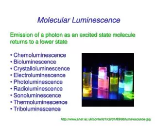
Luminescence • Emission of photons accompanying the relaxation from an excited to a ground state. • Photoluminescence—Excited state generated by absorption of a photon. • Fluorescence and phosphorescence • Chemiluminescence—Chemical reaction generates excited state. • Luminescence methods have greater inherent sensitivity than absorbance and often have greater linear dynamic range. • Disadvantage is that not all molecules luminesce and matrix interferences are more significant.
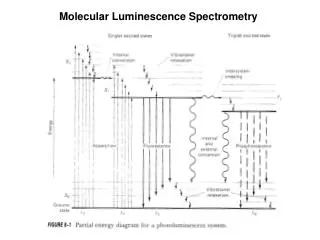
Fluorescence involves emission from states having the same spin. Lifetime 10-8-10-5sec Phosphorescence comes from “spin forbidden” transitions. Lifetime longer than 10-5 sec Seconds, minutes, hours Emission maximum of fluorescence and phosphorescence typically at lower energy than excitation radiation—Stokes shift. Exception: resonant emission (atomic fluorescence) What’s spin got to do with it?
Molecular energy level diagramsaka Jablonski diagrams fluorescence > absorbance phosphorescence > fluorescence
Quantum yield • A metric that describes efficiency of the fluorescent or phosphorescent process. • Approaches 1 for highly luminescent molecules • 0 for non-luminescent molecules • Ratio of the number of luminescent molecules compared to the total number excited. • Consider all deactivation pathways kf = fluorescent rate constant ki = intersystem crossing rate constant kec = external conversion rate constant kic = internal conversion rate constant kpd = predissociation rate constant kd = dissocation rate constant
Transition type and effects on fluorescence and phosphorescence • s*-s transitions rarely result in luminescence • Too high of energy (l < 250 nm) leads to predissociation and dissociation • Emission more common from p*-p, but also p*-n • From the lowest excited state • p*-p usually has greater quantum efficiency • Greater molar absorptivity of p-p* (10-100x) means high transition probability—short lifetime leads to large kf
Structural considerations • Fluorescence common in aromatic compounds with low-energy *- transitions • Conjugation shifts emission to red and greatly increases f • Example: pyridine vs. quinoline • Sensitive to substituents • Wavelengths of maximum absorption and emission and quantum yield • Halogens lead to sharp decreases in f • Heavy atom effect promotes intersystem crossing via spin-orbit coupling • Electronegativity also can give easily broken bonds • Structural rigidity enhances fluorescence • Lack of rigidity promotes non-radiative decay pathways (kic) • Example: Fluorene vs. biphenyl vs.
Environmental effects on fluorescence • Temperature • Increased number of collisions promotes external conversion. • Solvent • Heavy atoms in solvent promote intersystem crossing. • pH • Differing protonation states lead to resonance structures that change excited state energies • Concentration • When too many chromophores, the radiant power decreases through the sample so that not all species have chance to absorb and thus emit.
Quenching of fluorescence and phosphorescence • Nonradiative energy transfer from excited states to other molecules. • Dynamic (collisional) quenching—external conversion • Collision of excited species and quencher dependent on diffusion • Temperature, viscosity, and quencher concentration-dependent • Dissolved O2 is efficient quencher—degas solutions • Static quenching: quencher complexes with ground state fluorophore to form ‘dark complex’ • Förster quenching • Not dependent on collisions—long-range effect • Dipole-dipole coupling—falls off as 1/(distance)6 • Basis for Fluorescence Resonance Energy Transfer (FRET)
Wild type Mutant Nature Rev. Molec. Cell Biol., 2002, 3, 906-918. FRET microscopy http://upload.wikimedia.org/wikipedia/commons/3/3a/FRET.PNG http://www.bphys.uni-linz.ac.at/bioph/res/icg/Bilder/fret_methJD.png http://www.moleculardevices.com/pages/MM-new/metamorph_applications.html
Excitation and emission spectra A B B quinine C A D C D
Instrumentation for fluorescence and phosphorescence Why? • Almost always are double-beam • To compensate for radiant power fluctuations • Right angle detection Why?
Components of fluorometers and spectrofluorometers • Sources • Most common: Low-pressure Hg vapor lamps • 254, 302, 313, 546, 578, 691, and 773 nm lines • High-pressure Xe lamps • Continuum from 300-1300 nm • Lasers: essential for research operation with small samples, remote sensing (collimation), and when highly monochromatic light needed • Wavelength selectors • Interference filters or absorption filters for fluorometers • Grating monochromators (usually two) in spectrofluorometers • Transducers • PMTs often used for high sensitivity and often cooled to reduce S/N • CCDs for multichannel data acquisition
Correction of source and transducer variations with wavelength • Sources don’t have uniform power at all wavelengths which can bias excitation and emission spectra. • Most instruments have a reference spectrum stored in computer memory that can be used for instrumental correction.
Pros and cons of photo-luminescent analytical methods • Photoluminescence methods are inherently more sensitive than absorbance-based measurements • Fluorescence and phosphorescence dependent upon incident power, but measured independently of P0. • Absorbance requires measurement of P and P0—cannot be measured independently. • Also gives greater dynamic range because power can be modulated accordingly. • Typically give linear calibration plots and have high selectivity. • Photoluminescent measurements have less precision and accuracy. • Flicker noise and drift of source. • Background fluorescence, scatter, or quenching by matrix.
Applications of photoluminescence • Inorganic species determination • Formation of fluorescent complex via chelation • Fluorescence quenching—most common • Organic and biochemical species via fluorescence • Valuable tool to characterize foods, pharmaceuticals, clinical samples and natural products • Phosphorimetric methods • Complementary to fluorescent methods • Highly phosphorescent molecules often have weak fluorescence and vice versa. • Potentially greater selectivity because triplet conversion is required, but more difficult measurement (collisional quenching at RT problem with longer lifetimes) • Laser-induced fluorescence for detection in liquid chromatography
Fluorecence lifetime gives added element of selectivity Reports on collisional deactivation and energy transfer rates proximal to the fluorophore. 10 microsecond to sub-nanosecond time scale Fluorescent lifetime measurements http://www.olympusfluoview.com/applications/flimintro.html http://www.oxysense.com/technology/article/how_it_works/
Fluorescence microscopy • Subcellular fluorescence imaging • Combined with recombinantly-expressed fluorophores (GFP, etc.—Roger Tsien) has revolutionized biology. http://micro.magnet.fsu.edu/primer/techniques/fluorescence/anatomy/fluoromicroanatomy.html http://www.rp-photonics.com/img/kahn_fl_image.jpg
Normal Severe Dysplasia 1.4ns 2.4ns Fluorescence lifetime imaging (FLIM) Cancer Research, 2005, 65, 8766-8773. http://nimmi.bme.duke.edu/flim.html
Chemiluminescence • Excited state that emits light generated via a chemical reaction. • Actual mechanism usually quite complicated • Bioluminescence • Firefly, sea pansy, jellyfish, etc. A + B C* + D C* C + hn http://www.lifesci.ucsb.edu/~biolum/organism/photo.html
Analytical applications of chemiluminescence • Typically highly sensitive because there is no other source of light noise • “Zero background” measurement • Very simple instrumentation • No wavelength selection is typically needed • Signal intensity monitored over time • Analysis of gases • Nitric oxides: NO and NO2 via reaction with O3 • Analysis of strong oxidants or species that can generate strong oxidants (enzymatically). http://en.wikipedia.org/wiki/Luminol http://www.kpl.com/images/WESTERN2.JPG Luminol: 5-Amino-2,3-dihydro-1,4-phthalazinedione