Download
1 / 21
210 likes | 394 Views
HUMAN ANATOMY. The Appendicular Skeleton Ch. 8. LATIN TERMS to know!. Pect = breast Pelv = basin Sutur = seam Meta = after, adjacent Articulus = come together, joint. Vert = turn; joint Endo = within Epi = upon Lamina = thin plate Condyle = knuckle Sym = together.

E N D
HUMAN ANATOMY The Appendicular Skeleton Ch. 8
LATIN TERMSto know! • Pect = breast • Pelv = basin • Sutur = seam • Meta = after, adjacent • Articulus = come together, joint • Vert = turn; joint • Endo = within • Epi= upon • Lamina = thin plate • Condyle = knuckle • Sym = together • Be familiar with all bones & regions identified in the following notes & all underlined regions of bones!
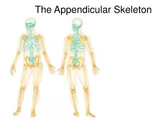
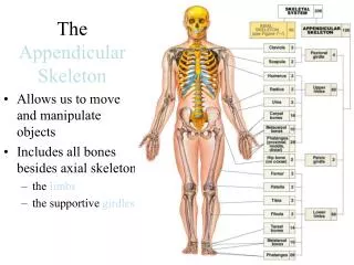
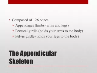
126 bones Consists of the: Upper Extremities Pectoral Girdle Humerus Ulna & radius Carpal bones Metacarpals Phalanges Lower Extremities Pelvic girdle Femur Tibia & fibula Tarsal bones Metatarsals Phalanges Appendicular Skeletal System
Pectoral girdle Clavicle Scapula Superior & medial borders Lateral border Inferior angle Caracoid process Acromion process Brachium Humerus Head Greater & lesser tuberosities Medial & lateral epicondyles Ulnar nerve Antebrachium Radius Ulna Olecranon process Trochlear (semilunar) notch Carpals Carpus Metacarpals Manus Phalanges Pollex The Upper ExtremitiesBones & bone regions to be familiar with!
SUPERIOR The Pectoral Girdle Manubrium • Clavicles • “S” shaped bones that originate at the superior lateral border of the manubrium of the sternum • Scapulae • Flat bones located at the posterior lateral portion of the body LATERAL MEDIAL INFERIOR
The Upper Limbs • Regions to be familiar with • Brachium (upper arm) contains the humerus • Antebrachium (forearm) contains the radius & ulna
Brachium • Humerus: Long bone that extends from the scapula to the elbow • Superior round portion that articulates with the scapula is known as the “head” • Greater& lessertuberosities • Medial&lateral epicondyles • Sites of skeletal muscle attachment • “Tuberosity” – refers to a process • “Epi” – on, “condyle” – knuckle • Ulnar nerve:runs the length of the humerus & attaches at the proximal end of the ulna (olecranon process) • Blow to this nerve sends sensation known as a “funny bone”
Antebrachium • The humerus articulates with the radius & ulna at a location known as the “condyle” • Ulna: long bone that is medial to radius • Olecranon process: superior/ proximal end of ulna • Forms point of elbow • Trochlear (semilunar) notch: large depression where distal end of humerus articulates with the olecranon process of the ulna • Radius: long bone that is the lateral bone of forearm
Wrist, Hand & Fingers Carpals Metacarpals Phalanges
Carpus & Manus Bones I V • Carpus (wrist) – • Contains 8 carpal short bones • 2 rows • Manus (hand) contains 19 bones in 2 groups • Metacarpals (5 in palm of hand) – • Short bones that articulate with distal carpal bones to support the hand • Roman numerals (I-V) are used to identify the metacarpals from lateral to medial • Phalanges (14 finger bones) – • Articulate distally to metacarpal bones • Proximal, middle & distal sets • Thumb is known as the pollex • Only has proximal & distal sets IV III II BRAIN BREAK: Is this diagram showing correct anatomical positioning?
The Lower ExtremitiesBones & bone regions to be familiar with! Pelvic girdle – ossa coxae Ilium Ischium Pubis Pubis symphysis Acetabulum Greater sciatic notch Ischial Tuberosity Femur Head Neck Shaft Greater (anterior) & Lesser (posterior) Trochanters Medial & Lateral Epicondyles Patella Fibula Head Tibia Tibial Tuberosity Lateral & medial condyles Tarsals Talus Calcaneous bone Navicular bone Cuboid Lateral, medial & intermediate cuneiform bones Metatarsals Phalanges
Pelvic Girdle • Paired hipbones - “ossacoxae” • Each hipbone – oscoxa • Fusion of 3 bones • Ilium (pl. – Ilia) • Extensive area of muscle, tendon, ligament attachment • Ischium(pl. – Ischia) • Posterior • Pubis • Anterior joint - Articulation of pubis bone at the anterior portion of the pelvic girdle – pubis symphysis • Fibrocartilage at joint • Posterior articulation – Ilia articulate to sacrum of vertebral column
More on the Ossa Coxae Ilium • Acetabulum • Articulation socket of ilia & head of each femur • All 3 bones of ossa coxae meet here • Greater sciatic notch – • Area through which large sciatic nerve runs & reaches lower extremities • Ischial Tuberosity – • Projection on posterior, lateral side of ischia • Bears all body weight when sitting Pubis Ischium
Comparison of Male & Female Pelvic Girdles • Female: • Less massive, shallower pubic arch, pelvic inlet round/oval • Male: • Heavier, upper pelvis nearly vertical, coccyx more vertical, pelvic inlet heart-shaped, outlet smaller
The Lower Limbs • Femur • Fibula • Tibia Is this person standing in correct anatomical position?
FEMUR • Longest & heaviest bone in body • Articulates proximally with ossa coxae at hip joint & distally with tibia at knee joint • Regions to identify: • Head • Neck • Shaft • Greater (anterior) & Lesser (posterior) Trochanters • Medial & Lateral Epicondyles
Patella (Kneecap) • Triangular sesamoid bone • Enclosed in the quadriceps tendon that secures the anterior thigh muscles to the tibia (lower limb) • Guards knee joint anteriorly & improves leverage of thigh muscles acting across knee joint
Tibia & Fibula • Tibia – • Large medial bone that articulates with the epicondyles of the femur • Helps support weight • “shinbone” • Fibula – • Parallels the lateral border of the tibia • Aids in moving foot & toes
This little piggy went to the market… I • Tarsal bones: • Talus: ankle • Calcaneous bone: heel bone • Navicular bone • Cuboid • Lateral, medial & intermediate cuneiform bones • Metatarsals – • Short bones that articulate with distal tarsal bones • Identified by Roman Numerals (I-V) • Phalanges (toes, digits) • Articulate distally to metatarsal bones • Proximal, middle, distal (14 total) • Big toe – “Great toe” • Has 2 phalanges (proximal & distal) II III IV V