Download
1 / 167
1.73k likes | 1.99k Views
14. Thalassemia. Learning Objectives—Level l. At the end of this unit of study, the student should be able to: Define thalassemia. Differentiate thalassemias from hemoglobinopathies based on definition and basic pathophysiology.

E N D
14 Thalassemia
Learning Objectives—Level l At the end of this unit of study, the student should be able to: • Define thalassemia. • Differentiate thalassemias from hemoglobinopathies based on definition and basic pathophysiology. • Describe the typical peripheral blood morphology associated with thalassemia. • Compare and contrast the etiology of α- and β-thalassemia. continued on next slide
Learning Objectives—Level l At the end of this unit of study, the student should be able to: • For each of the four genotypes of α-thalassemia, describe the: • Number of affected alleles • Individuals affected • Basic pathophysiology continued on next slide
Learning Objectives—Level l At the end of this unit of study, the student should be able to: • For each of the four genotypes of α-thalassemia, describe the: • Symptoms • Laboratory results including blood cell morphology and hemoglobin electrophoresis continued on next slide
Learning Objectives—Level l At the end of this unit of study, the student should be able to: • For each of the six genotypes of β-thalassemia describe the: • Individuals affected • Basic pathophysiology • Symptoms • Laboratory results including blood cell morphology and hemoglobin electrophoresis
Learning Objectives—Level ll At the end of this unit of study, the student should be able to: • List and describe five primary genetic defects found in thalassemias. • Compare and contrast α- and β-thalassemia. • Correlate the outcomes in hemoglobin synthesis resulting from the five genetic defects in thalassemia.
Learning Objectives—Level ll At the end of this unit of study, the student should be able to: • For all four genotypes of α-thalassemia: • Correlate all three nomenclature systems: genotype, genotype description, and phenotype. • Explain advanced pathophysiology. • Describe treatment and prognosis. continued on next slide
Learning Objectives—Level ll At the end of this unit of study, the student should be able to: • For all four phenotypes of β-thalassemia: • List expected genotypes. • Explain advanced pathophysiology. • Describe treatment and prognosis. • Correlate clinical severities of both α-thalassemia and β-thalassemia with their respective genotypes. continued on next slide
Learning Objectives—Level ll At the end of this unit of study, the student should be able to: • Compare and contrast other thalassemia and thalassemia-like conditions to include: • δβ-thalassemia • γδβ-thalassemia • Hemoglobin Constant Spring • Hereditary persistence of fetal hemoglobin (HPFH) continued on next slide
Learning Objectives—Level ll At the end of this unit of study, the student should be able to: • Compare and contrast other thalassemia and thalassemia-like conditions to include: • Hemoglobin Lepore • Thalassemia/hemoglobinopathy combination disorders continued on next slide
Learning Objectives – Level ll At the end of this unit of study, the student should be able to: • Differentiate iron-deficiency anemia and HPFH from thalassemia based on results of laboratory tests and clinical findings.
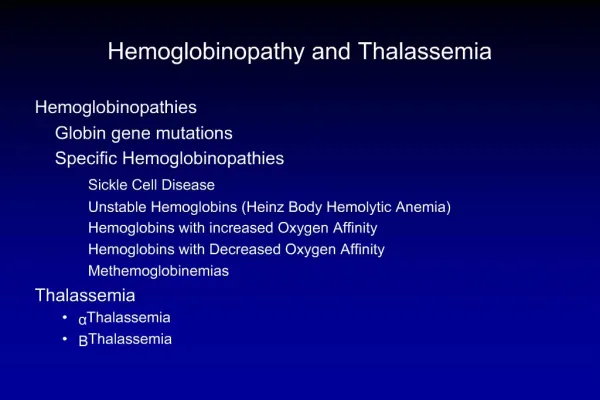
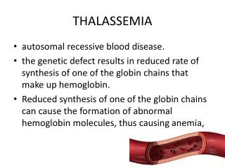
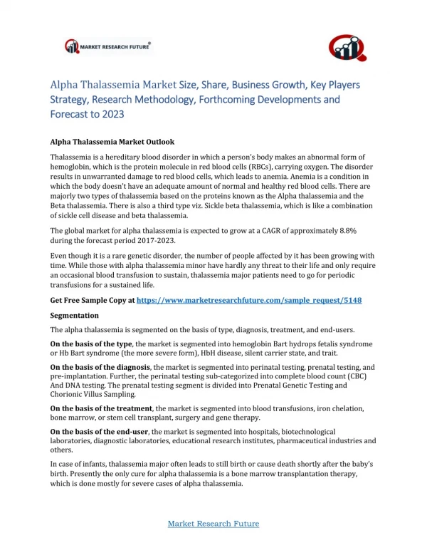
Introduction • Inherited disorders • Mutations in one or more globin genes • Decreased or absent synthesis of globin chain(s) • > 400 unique mutations • α-thalassemia • β-thalassemia
Introduction • Thalassemia is one of the most common genetic disorders • Major health problem
Thalassemia vs. Hemoglobinopathy • Hemoglobinopathy • Qualitative defects • Production of abnormal Hb molecules • Usually point mutations
Thalassemia vs. Hemoglobinopathy • Thalassemia • Quantitative disorders • Produce reduced amounts of normal Hb • Both deletional and nondeletional mutations • Reduced amounts of normal Hb
Table 14-1 Comparison of Hemoglobinopathies and Thalassemias
Genetic Defects in Thalassemia • Five categories of genetic defects • Gene deletion • Promoter mutation • Nonsense mutation • Mutated termination (stop) codon • Splice site mutation continued on next slide
Genetic Defects in Thalassemia • Results in the mutated globin chain • Absent • Reduced in concentration • Longer or shorter than normal
Figure 14-2 Hemoglobin electrophoresis on cellulose acetate or agarose at pH 8.4 is helpful in distinguishing the type of thalassemia and in differentiating thalassemias from hemoglobinopathies. In -thalassemias, there is a reduction in -containing hemoglobins (HbA, HbA2, and HbF) proportional to the number of deleted -genes and in the more severe cases, the emergence of non--containing hemoglobins (HbH and Hb Bart’s).
Types of Thalassemia • Six versions of thalassemia • α, β, γ, δ, ε, ζ • Normal adult globin chains • α, β, δ, γ • 97% of normal adult Hb is HbA (α2, β2) • Deficiency of α- or β-chains affects HbA • Reduces blood's O2 carrying capacity and HbA concentration
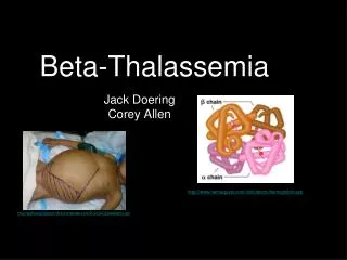
Types of Thalassemia • Two major types of classical thalassemia • α-thalassemia • Impaired α-chain synthesis • β-thalassemia • Impaired β-chain synthesis
Types of Thalassemia • Rare thalassemia • δ-thalassemia • Not clinically significant, rare • Combinations of gene deletions • δ β, γδβ, rare
Types of Thalassemia • Occasionally structural Hb variant • Decreases globin chain synthesis • Clinical picture of thalassemia • Hbs with abnormally long or short globin chains Ex: Hb Constant Spring • HbE—point mutations continued on next slide
Types of Thalassemia • Occasionally structural Hb variant • Hb Lepore—structural variant and ineffective synthesis • Hereditary persistence of fetal Hb (HPFH) • Variant of β-thalassemia, ↑ Hb F production
Pathophysiology • Normal ratio of α- and β-chains is 1.0 • ↓ or absent synthesis of one chain results in excess of other chain • Imbalance contributes to • ↓ total RBC Hb production • Ineffective erythropoiesis • Chronic hemolysis
Pathophysiology • Excess α-chains • Highly insoluble • Precipitate in the cell • Bind to cell membrane causing damage • ↓ RBC deformability • BM macrophages destroy precipitate-filled RBCs causing ineffective erythropoiesis continued on next slide
Pathophysiology • Excess α-chains • Circulating RBCs with precipitates • Pitted or removed by the spleen • Chronic extravascular hemolysis
Pathophysiology • Excess β-chains • Combine to form HbH (β4) • High O2 affinity • Unstable • Excess γ-chains • Fetus • Hb Bart's γ4 • High O2 affinity
Clinical Findings • Anemia caused by: • Decreased HbA synthesis • Chronic hemolysis • Ineffective erythropoiesis • Severity depends on: • Specific genetic mutation • Number of genes affected
Clinical Findings • Chronic hemolysis • Splenomegaly • Spleen is major site of extravascular hemolysis • Functional hyposplenism • Overburdened by RBC destruction • Gallstones • Formed from large amounts of bilirubin excreted by liver
Clinical Findings • Chronic demand for RBCs • BM increases erythropoiesis • BM expansion and thinning of calcified bones • Skeletal abnormalities • Pathologic fractures continued on next slide
Clinical Findings • Chronic demand for RBCs • Increased iron absorption causes • Iron toxicity • Extramedullary erythropoiesis • Compression syndromes
Clinical Findings • Pregnant women with thalassemia • Developing infants impacted more than mother • Diminished growth • Premature birth • Intrauterine death
Laboratory Findings • Peripheral blood • Microcytic hypochromic anemia • ↓ MCV, MCH, MCHC • RBC count normal or ↓, but ↑ relative to hemoglobin and hematocrit levels • RDW may or may not be ↑ continued on next slide
Laboratory Findings • Peripheral blood • Target cells, basophilic stippling, NRBCs • Precipitated excess chains • Seen on supravital stain • Reticulocyte ↑ • Bilirubin ↑, haptoglobin ↓
Laboratory Findings • Hb electrophoresis • HbA ↓ • β-thalassemia • HbF ↑, HbA2 ↑ • α-thalassemia • HbF ↓, HbA2 ↓ • Hb Bart's and HbH present
Laboratory Findings • BM (not necessary for diagnosis) • Erythroid hyperplasia • Erythroblasts abnormal • ↓ cytoplasm • Striking basophilic stippling • ↑ iron
Laboratory Findings • Screening tests • Allelle-specific oligonucleotide hybridization • Dot blot and reverse dot blot assays • Amplification refractory mutation system • Direct sequencing • Nucleic acid based methods • For prenatal diagnosis
Table 14-3 Clinical and Laboratory Findings Associated with Thalassemia
α-Thalassemia • Etiology • α-thalassemia is group of four disorders characterized by ↓ synthesis of α chains • Two α-genes on each of two #16 chromosomes = fourα-genes (diploid) continued on next slide
α-Thalassemia • Etiology • Four clinical severities • All four α-genes deleted • Hydrops fetalis • Three of the four α-genes deleted • HbH disease continued on next slide
α-Thalassemia • Etiology • Four clinical severities • Two of the four α-genes • α-thalassemia minor • One of the four α-genes • Silent carrier
α-Thalassemia • Affected alleles • α-chains produced proportional to the number of affected alleles • Four α-chains • Two pairs, α1 andα2 • α2-gene produces 2–3× more mRNA than α1-gene • α2-gene deletion more severe than α1-gene continued on next slide
α-Thalassemia • Affected alleles • α-chains produced proportional to the number of affected alleles • Internal mechanism • Stimulate ↑ production of α-chains from unaffected genes to compensate for deletions
α-Thalassemia • Affected individuals • Found primarily in people of Mediterranean, Asian, African ancestry • Commonly seen in blacks, Indians, Chinese, Middle Eastern people • Patients from African descent • Milder version of α-thalassemia • Usually involves α1-gene (lower producing gene)
Figure 14-1 A short section of chromosome 16 showing the 5 to 3 orientation of three functional genes , 2, and 1 along with three pseudogenes , 2, and 1. Pseudogenes are the result of partial gene duplications but are not expressed. There are two functional -genes on each chromosome; the 2-gene expresses 2–3 times as much protein product as the 1-gene.
α-Thalassemia • Genotypes • Three nomenclature systems • Genotypic, genotypic description, phenotypic • Genotypic system • Deleted genes (–) • Unaffected genes (α) continued on next slide
α-Thalassemia • Genotypes • Genotypic description system • α-thal-1 or α0 • Deletion of both α genes on the same chromosome (–,–) • α-thal-2 or α+ • One gene deleted, one normal gene on the same chromosome (–,α)
α-Thalassemia • Phenotypic system • Describes four clinical types • Hydrops fetalis • HbH disease • α-thal minor • Silent carrier