Download
1 / 32
340 likes | 658 Views
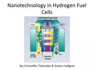
Mast cells & basophils I: Development & function. Feb. 23, 2004 Advanced Immunology Course Stephen J. Galli (sgalli@stanford.edu) Janet Kalesnikoff (jkalesni@stanford.edu). The setting: Parasite immunity and allergic diseases

E N D
Mast cells & basophils I: Development & function Feb. 23, 2004 Advanced Immunology Course Stephen J. Galli (sgalli@stanford.edu) Janet Kalesnikoff (jkalesni@stanford.edu)
The setting: Parasite immunity and allergic diseases • Mast cell and basophil development (and their phenotypic heterogeity) • Basic aspects of function: Regulation of FceRI expression; secreted products • Positive and negative regulation of activation via the FceRI (Janet Kalesnikoff) • Other activation mechansims; proposed roles in health & disease
Asthma • Affects approximately • 7% of population in U.S. • Features • Reversible airway narrowing • Immunologically non-specific airway hyperresponsiveness (AHR) • Chronic inflammation of the airways • Airway remodeling
Asthma • PREVALENCE INCREASED 75% FROM 1980 TO 1994 • > 1.8 MILLION EMERGENCY ROOM VISITS IN 1995 • > 5,300 DEATHS FROM ASTHMA ANNUALLY • DIRECT COSTS > $12.6 BILLION ANNUALLY • INDIRECT COSTS > $2.8 BILLION ANNUALLY • > 10 MILLION SCHOOL DAYS MISSED ANNUALLY
Potential roles of mast cell products in atopic asthma C.M.M. Williams & S.J. Galli. J. Allergy. Clin. Immunol. 105:847, 2000.
Mast cell & basophil development, heterogeneity & function: Mast cells & basophils are not the same T. Kawakami & S.J. Galli. Nature Rev Immunol. 2: 773, 2002. Reprinted with permission from A. Dvorak et al.Hum. Pathol. 11:606, 1980.
Highly simplified diagram of mast cell & basophil development (in the mouse)
Hematopoiesis: the formation of blood cells Lymphoid Stem Cell Pluripotent Stem Cell Myeloid Stem Cell Mast cell
Figure 1: A tentative scheme of hematopoiesis and its growth factors. The pluripotent stem cell divides infrequently to generate either more stem cells (self-renewal; as indicated by ) or committed progenitor cells which can produce only one or a few types of blood cells. Progenitor cells are stimulated to proliferate and differentiate by specific growth factors (indicated on the arrows) but progressively lose their capacity for division and develop into terminally differentiated blood cells, which live for only a few days or weeks. Only mature T cells, B cells, mast cells and macrophages are known to carry proliferative potential (as indicated by ). Dotted arrows represent uncertain pathways. Transcription factors involved in the differentiation of specfic pathwas are indicated in italics under the arrows. SCF = stem cell factor; IL = interleukin; GM-CSF = granulocyte monocyte colony stimulating factor; G-CSF = granulocyte colony stimulating factor; M-CSF = monocyte colony stimulating factor; TPO = thrombopoietin; EPO = erythropoietin. CFU = colony forming unit. BFU = burst forming unit. Adapted from Alberts et al.4 SCF IL-2 NK cell NK precursor IL-2 IL-7 IL-7 flk-2/flt-3 ligand SCF flk-2/flt-3 ligand SCF GATA-3 T cell Pre-T cell IL-8 flk-2/flt-3 ligand SCF Lymphoid stem cell IL-7 flk-2/flt-3 ligand SCF PU.1 Pax5 E2A Ikaros PU.1 Pre-B cell B cell SCF IL-3 Pluripotent stem cell flk-2/flt-3 ligand SCF SCF IL-3 GATA-1/2 Elf-1 SCF IL-3 CFU-mast Mast cell PU.1 C/EBP SCF IL-3 IL-3 Myeloid stem cell SCF IL-3 CFU-Bas Basophil IL-3 GM-CSF SCF IL-3 flk-2/flt-3 ligand GATA-1 C/EBP Ets-1 Eosinophil CFU-Eos IL-3 GM-CSF G-CSF TPO SCF IL-3 PU.1 C/EBP Neutrophil IL-3 GM-CSF M-CSF GM-CSF M-CSF CFU-GM SCF IL-3 PU.1 PU.1 Macrophage SCF IL-3 GM-CSF IL-11 IL-6 TPO Monocyte Osteoclast Platelets GATA-1 FOG NF-E2 GATA-1/2 CFU-Meg Megakaryocyte SCF IL-3 GM-CSF EPO IL-3 GM-CSF EPO GATA-1 GATA-1 NF-E2 EKLF FOG Erythrocyte BFU-E CFU-E
Ching-Cheng Chen (C3), Devavani Chatterjea (DC), et al. Murine mast cell/basophil development Define lineage relationships of mast cells (C3)/basophils (DC) to other hematopoietic cells. Pro-B B CLP NK Pro-T LT-HSC T ST-HSC MPP DC GMP Mf GR CMP MEP PL E ? MC
T. Kawakami & S.J. Galli. Nature Rev Immunol. 2: 773, 2002. FceRI-mediated activation pathways in mast cells
1) Preformed, granule-associated proteases, proteoglycans (e.g., heparin) and bioactive amines (e.g. histamine); released by DEGRANULATION 2) Newly synthesized AA metabolites (e.g. LTC4 & PGD2) 3) Diverse cytokines; chemokines (e.g. TNF, IL-4, IL-6; MIP-1, IL-8) LTC4 PGD2 IL-4, IL-5, IL-6, IL-8, IL-13, TNF, MIP-1, etc. Activated mast cells/basophils release 3 major classes of “pro-inflammatory” mediators* * Note: Underlined products are secreted by mast cells & basophils; basophils secrete a much more restricted set of cytokines than do mast cells.
IgE-dependent enhancement of FceRI surface expression: Functional consequences • As surface expression of FceRI increases, mast cells: • Can be “sensitized” to more antigens (Ags) • Can release mediators at lower Ag concentrations • Can release larger amounts of products in response to Ag • May release additional products (e.g., IL-4 in mouse mast cells) in response to Ag challenge Reviewed in: C.M.M. Williams & S.J. Galli. J. Allergy. Clin. Immunol. 105:847, 2000.
cDNA microarray analysis of human mast cells stimulated via aggregation of FceRI * • Generate human umbilical cord blood-derived mast cells of >98-99% purity in SCF and IL-6. • Sensitize mast cells with human myeloma IgE (5 mg/ml) and rhIL-4 (10 ng/ml) for 4 days [to increase FceRI on cell surface]. • Challenge mast cells with anti-human IgE (10 mg/ml). • Assess % histamine release at 1 h; recover mRNA for microarray studies at baseline (0 hr), 1 & 2 h; measure released cytokines at various time intervals. * K. Sayama, M. Diehn, K. Matsuda, C. Lunderius, M. Tsai, S-Y. Tam, D. Botstein, P.O. Brown & S.J. Galli. BMC Immunol. 3:5, 2002.
Clustering of > 2,400 genes in two human mast cell populations (> 98% pure, cord blood-derived) stimulated via FcRI Donor 1 Donor 2 0 1h 2h 0 1h 2h Histamine Release (%) at 1 h Vehicle Anti-IgE Donor 1 2 ± 0.1 57 ± 4 Donor 2 3 ± 0.2 63 ± 2 • 2,478 genes of ~ 14,000 tested exhibited 2-200 fold changes in expression vs. baseline (time 0) • ~ 50% increase(red) • ~ 50% decrease(green) • ~ 50% of these 2,478 genes represented ESTs of genes of unknown function “Reference” mRNA from: T cells (Jurkat), B cells (Raji), mast cells (HMC-1) [80% “resting”: 20% 2h with 1 mM ionomycin, 25 ng/ml PMA] K. Sayama, M. Diehn, K. Matsuda, C. Lunderius, M. Tsai, S.-Y. Tam, D. Botstein, P.O. Brown & S.J. Galli. BMC Immunol. 3:5, 2002.
Cytokines Chemokines Donor 1 Donor 2 0 1h 2h 0 1h 2h Donor 1 Donor 2 IL-16 † IL-1 IL-14 * MIF TNF- converting IL-11 *enzyme * IL-9 IL-6 IL-5 IL-4 CSF-1 IL-18 IL-12 p40 † TNF- IL-3 Pre-B cell enhancingfactor * GM-CSF IL-1 LIF † 0 1h 2h 0 1h 2h ‡ Eotaxin Gro2 IL-8 MCP-1 HCC-4 * MIP-1 RANTES LD78 * MIP-1 MCP-3 Lymphotactin MIP-3 * MCP-4 * ‡ ‡ ‡ * = “new” for any mast cells ‡ = “new” for human mast cells † = “new” information about FceRI-induced change in expression K. Sayama, M. Diehn, K. Matsuda, C. Lunderius, M. Tsai, S.-Y. Tam, D. Botstein, P.O. Brown & S.J. Galli. BMC Immunol. 3:5, 2002.
CD40 ligand † CD82 † SLAM * Lymphotoxin- * ICAM-1 † CD83 * 4-1BB ligand * OX40 ligand * Gene products that regulate cell-cell interactions between mast cells & other cell types Adhesion Molecules T Cell, B Cell and Dendritic Cell Interaction Molecules Donor 1 Donor 2 Donor 1 Donor 2 0 1h 2h 0 1h 2h 0 1h 2h 0 1h 2h P-selectin glycoprotein CD103* ligand † CD49F † ELAM-1 * CD49B † CD29 † ICAM-1 † Protocadherin 43 * K. Sayama, M. Diehn, K. Matsuda, C. Lunderius, M. Tsai, S.-Y. Tam, D. Botstein, P.O. Brown & S.J. Galli. BMC Immunol. 3:5, 2002.
Crosslink with multivalent Ag Lyn Fyn Fyn Lyn Blk & Fyn Lck • Associated with Src family tyrosine kinases phosphorylate ITAM motifs (green) serve as docking sites for adaptors or other NRTKs • Immunoreceptors/antigen receptors: multiple subunits TCR BCR FcRI MHC I/II CD4/8 peptide surface bound Ig CD3
IgE Antigen- binding sites • 5 Classes of immunoglobulins or antibodies - IgG, IgM, IgA, IgD and IgE • IgE heavy chain: -chain • Binds high affinity FcRI Fab FC FcRI • Antigen receptor superfamily • Tetrameric structure • 3 Distinct protein species - binds to Fc portion of IgE - 4 transmembrane domains - 1 transmembrane domain - disulfide linked homodimer signal transduction
IgE CURRENT IgE/MAST CELL DOGMA • IgE binds to mast cells with high affinity (via FcRI) • IgE binding is a “passive sensitization” step • Activation occurs only when bound IgE is cross- linked, e.g., by binding to a multivalent Ag
DNP HSA DNP DNP LAT LAT GTP Btk PDK1 Gab2 Ras Syk Lyn PI3K Fyn Fyn Sos Grb2 Slp-76 Raf-1 Vav Akt Rac DAG + IP3 MEK p38 JNK [Ca2+] PKC ERK PLA2 IgE & Ag-induced mast cell activation IgE IgE FceRI FceRI PI-4,5 -P2 PI-3,4,5-P3 PLC Degranulation Cytokine/chemokine production Arachidonic acid metabolism (LTC4, PGD2)
DNP HSA DNP DNP FcRIIB Crosslink with Ag IgG Gab2 Syk Lyn Fyn PI-3,4,5-P3 PI-4,5 -P2 PI-3,4-P2 LAT Btk PDK1 Fyn PI3K SHIP PLC Dok DAG + IP3 Akt RasGAP SHIP [Ca2+] PKC Shc Ras Negative regulation of IgE & Ag-induced mast cell activation IRS (inhibitory receptor superfamily) • Contain 1+ ITIM motif (red) • Coaggregation with FcRI inhibits signalling FcRI IgE IgE
IRS (inhibitory receptor superfamily) # ITIMs Recruits Ligand FcRIIB 1 SHIP IgG+Ag gp49B1 2 SHP-1 v3 PIR-B 4 SHP-1 SHP-2 SHIP ? SIRP 4 SHP-1 SHP-2 CD47 MAFA 1 SHIP ? H. R.Katz (2002). Curr Opin Immunol 14:698-704.
DNP HSA DNP DNP PI-3,4-P2 PI-3,4,5-P3 LAT LAT SHIP SHIP2 GTP Gab2 Ras Syk Lyn Lyn Btk PI3K Fyn Fyn Sos Grb2 PLC PDK1 Slp-76 Raf-1 SHP1 Vav Rac Akt DAG + IP3 MEK p38 JNK ERK [Ca2+] PKC PLA2 Arachidonic acid metabolism (LTC4, PGD2) Cytokine/chemokine production Degranulation Negative regulation of IgE & Ag-induced mast cell activation Intracellular/cytoplasmic inhibitors IgE IgE FceRI FceRI PI-4,5 -P2 RasGAPs Rin1 RabGEF1?
RabGEF1 is a negative regulator of mast cell activation and skin inflammation See-Ying Tam, Mindy Tsai, John N. Snouwaert, Didier Scherrer, Devavani Chatterjea, Donna M. Bouley & Stephen J. Galli. Nature Immunology (submitted). RabGEF1 knockout mouse
5M3+/+ 5M5-/- 20 ng/ml DNP IgE & Ag-induced Ca2+ flux is elevated in RabGEF1-/- BMCMCs Fluo-4/Snarf-1
IgE & Ag-induced release of pre-formed mediators is elevated in RabGEF1-/- BMCMCs +/+ +/- -/- % Degranulation 1ng/ml DNP 10ng/ml DNP 100ng/ml DNP 1g/ml DNP - A&P
IgE & Ag-induced LTC4, PGD2 and cytokine production are elevated in RabGEF1-/- BMCMCs
Stimulus PI-3,4,5-P3 PI-4,5-P2 DAG GTP Shc Ras PI3K PLC PKC Sos Rab5 Ca2+ PKB Btk IP3 Raf-1 Early Endosome RabGEF1 ? Cell survival MEK Endocytosis ERK Mitogenesis
Diverse potential mast cell activation mechanisms in host defense/pathology * • Products of pathogens (LPS/CD14-TLR4, E. coli FimH/CD48, C. difficile toxin, H. pylori, etc., etc.) • Products of complement activation (C3a/C3aR, C5a/C5aR, ? C3bR/CD35, etc.) • IgE (FceRI, CD23, galectin-3), IgG1 (FcgRIII, mouse), LCvia antigens or superAgs (e.g., S. aureus Protein A, P. magnus Protein L, HIV gp120, protein Fv in HBV & HCV) • T cell-derived products (?) • Other: neuropeptides (e.g., VIP, Neurotensin, Substance P), ET-1, SCF & other cytokines, leukocyte products, defensins/LL-37, insect/reptile venom components, etc., etc. * Responsiveness (and responses) of different mast cell (basophil) populations can vary: “Mast cell (basophil) heterogeneity”
Effector (& immunoregulatory) cell in: IgE- &/or (in mice) IgG1-associated immune responses, e.g., anaphylaxis, “asthma” & parasite immunity; some mouse models of “autoimmunity” (e.g., MS, RA) Proposed mast cell (basophil ?) roles (a very incomplete list-I) • Regulate “immunologically non-specific” acute and chronic inflammation & “natural immunity” (e.g., IBD) • Regulate wound healing, angiogenesis & tissue-remodeling • Regulate T cell-dependent, “Ig-independent” responses (e.g., CS)
Promote &/or retard tumor development, progression or metastasis Bi-directional interactions with peripheral nerves & promotion of “neurogenic inflammation” & neurite growth Proposed mast cell (basophil?) roles (a very incomplete list-II) • Promote protective responses to diverse endogenous or exogenous noxious (non-microbial) agents • Regulate epithelial development, proliferation & function (e.g., “barrier function”, hair growth)