Download
1 / 61
610 likes | 804 Views
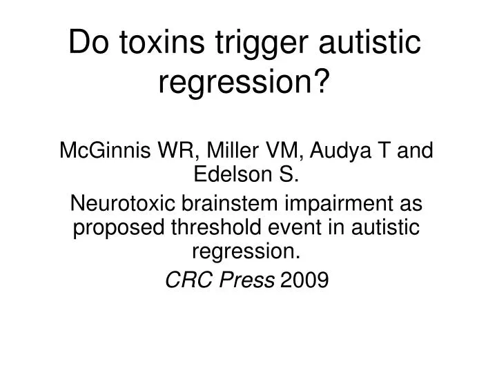
Do toxins trigger autistic regression?. McGinnis WR, Miller VM, Audya T and Edelson S. Neurotoxic brainstem impairment as proposed threshold event in autistic regression. CRC Press 2009. Intriguing phenomenon of autistic regression. Widely recognized Usually 18-24 months of age

E N D
Do toxins trigger autistic regression? McGinnis WR, Miller VM, Audya T and Edelson S. Neurotoxic brainstem impairment as proposed threshold event in autistic regression. CRC Press 2009
Intriguing phenomenon of autistic regression • Widely recognized • Usually 18-24 months of age • Relatively rapid: days or weeks • Earlier problems in some children • Published incidence as high as 50%
Features of regression • Vocalization loss: acquired words or babbling (29% / 9%. Lord 2004) • Loss of social function, in some cases unassociated with loss of vocalization (Goldberg 2003) • Gastrointestinal impairment (Madsen 2004; Goldberg 2004)
GI tract in regressed cohorts • Radiographic fecal loading or megacolon (100%. Torrente 2002) • Reflux esophagitis (69%. Horvath 1999) • Enterocolitis (88%. Wakefield 1998, 2002)
Toxins are plausible triggers • Parallel increase in autism and environmental toxicants (Lathe 2008) • Autism rates correlate with: 1. Presence of toxic landfills (Ming 2008) 2. Estimated environmental cadmium and mercury (Windham 2006) 3. Proximity to mercury point sources (Palmer 2009)
Toxins as triggers • Elevated dental concentrations of mercury (Adams 2007) • Autistic symptoms correlate with mercury-consistent porphyrins (Geier 2008) • Case reports: Geier: temporal association with mercury by injection Bradstreet, et al: on-off speech on DMSA
The case of R.K. • Progressive loss of speech over weeks post one-time placement of 9 amalgams at age 4. • By age 6, had regained 200 words, but elevated blood [Hg] explicable only on basis of amalgams. • Subsequent loss of all speech immediately after one-time removal of amalgams without precautions.
Toxins as triggers • Many neurotoxins are oxidative, and oxidative modification is increased in brain of autistic children (Evans 2008; Lopez-Hurtado 2008; Sajdel-Sulkowska 2008) • Gliosis and neuronal loss in autism is consistent with toxicant effects (Kern 2006)
Circumventricular Organs (CVO) Area postrema (AP) Posterior pituitary Median eminence (ME) Subfornical Organ Organum vasculosum Pineal Gland [Nucleus Tractus Solitarius (NTS)] • Portals for toxins: no blood-brain barrier (BBB) • In and around the brainstem
CVO are preferentially sensitive to a broad class of neurotoxins • Cadmium • Monosodium glutamate (MSG) • Paraquat • Inorganic mercury (including inorganic mercury from metabolic conversion of organic and elemental forms)
Cadmium • Injections accumulate only in brain outside BBB, including AP and pineal (Arvidson 1986) • Lipid peroxidation (Mendez-Armenta 2003) blocked by antioxidants (Kim 2008) • Inhibits complex II and III (Wang 2004)
MSG • Injections accumulate only in brain outside BBB, including AP (Karcsu 1985) and ME (Meister 1989; Peruzzo 2000) • Lipid peroxidation, persistent for long periods (Bawari 1995; Singh 2003) • Excitotoxic. In autism, GAD much lower in brain (Fatemi 2002)
Paraquat • Injections accumulate only in areas outside the BBB, including AP and pineal (Naylor 1995) • Increased TNF-α and superoxide production by microglia (Wu 2005) • Inhibits complexes II and IV (Palmeira 1995)
Inorganic mercury • Worrisome levels in air, water, soil (McGinnis 2001) and food (Dufault 2009) • Pink Disease proves fractional systemic absorption in children (McGinnis 2001) • Injections accumulate in AP and brainstem motor nuclei (Arvidson 1992) • Persists in brain for years (Vahter 1994) • Immune stimulation (Havarinasab 2007) and increased microglia (Geier 2007)
Elemental mercury Amalgam removal decreased plasma and red-cell inorganic mercury levels by 73% (Halbach 2008)
Daily oral organic (methyl) mercury in primates • In 6 brain areas, inorganic mercury averaged x30 at 6 mos., x60 at 18 mos. • By far, highest inorganic mercury at pituitary, only CVO examined. • If stop organic dosing at 6 mos,inorganic mercury doubles in pituitary at 12 months, but not in regions with BBB. (Vahter 1994)
Brainstem • The “hard-drive” control panel for messages between brain and body • Rimland emphasized brainstem in 1964, but scant current neuropath interest • Many findings in autism consistent with brainstem dysfunction
Brainstem abnormalities • Smaller medulla and midbrain on MRI (Courchesne 1997; Hashimoto 1993) • Reduced gray matter on MRI (Jou 2008) • Ectopic neurons and aberrant tracts (Bailey 1998) • Swollen medullary, thalamic, hypothalamic axon terminals (Weidenheim 2001) • Abnormalities of inferior and superior olives (Kemper 1993; Kulesza 2008)
Brainstem abnormalities • Auditory brainstem response (Klin 1993; Kwon 2007) • Centrencephalic EEG (Gilberg 1983) • Heart rate, respiratory and vascular response (Bonvallet 1963; Althaus 2004) • Post-rotatory response (Ornitz 1983)
Suggest CVO impairment • Pineal: abnormal melatonin production (Nir 1995; Kulman 2000; Tordjmann 2005) • Median eminence / posterior pituitary: abnormal oxytocin production (Modahl 1998; Green 2001)
Dorsal vagal complex (DVC) • Area postrema (AP) • Dorsal motor nucleus of vagus (DMV) • Nucleus tractus solitarius (NTS) The DVC mediates autonomic function of cervical, thoracic and abdominal viscera.
Area postrema • No BBB • Highly vascular and very long residence time of blood in capillaries • So-called “emesis center” • Ablation increases consumption of water or concentrated salt water, food aversions, craving for carbohydrates and bland food • [Flavor aversion 2° Cd reversed by DMSA]
Nucleus tractus solitarius • Lacks BBB on one side (Gross 1990) • Viscerosensory and visceromotor; parasympathetic and sympathetic efferent • Mediates social behavior, arousal, mood, emotion, anxiety, seizure activity and pain via limbic and cortical projections (Marvel 2004; Nemeroff 2006)
Secretin and NTS • Highest binding of infused secretin at NTS; secretin activates NTS neurons (Yang 2004) • Several studies reported improvements in social behavior—may relate to NTS effect (Myers 2008) • Parent reports of sudden potty-training consistent with NTS effect (Beck, et al.)
Dorsal motor nucleus of the vagus (DMV) • Visceromotor: peristalsis, esophageal sphincter tone, heart rate, pharyngeal and laryngeal musculature • Tensor palati to open eustachian tube • Viscerosecretory: digestion, floral balance
Phonation • Significantly autonomic, “subconscious” • DMV visceromotor to all the intrinsic and extrinsic muscles of larynx and pharynx • Vagal alteration results in altered pitch (Shaffer 2005) • Vagal dysarthria should not be confused with classical “motor apraxia of speech”
Suggest DVC impairment • Excessive thirst (Terai 1999) • Salt-craving and flavor-aversion (ARI) • Otitis media (Konstantareas 1987; Rosenhall 1999) • Abnormal heart rate, respiratory and vascular (Bonvallet 1963; Althaus 2004) • Depressed cardiac parasympathetic and abnormal baroreflex (Ming 2005)
Suggest DVC impairment • Esophageal reflux in 67% of regressed cohort (Horvath 1999) • Fecal loading or megacolon in 100% of regressed cohort (Torrente 2002) • Enterocolitis in 88% of regressed cohort (Wakefield 1998) • Paneth’s cells enlarged with granules (Horvath 1999)
Duodenal Paneth’s cells (Courtesy of K. Horvath, Thomas Jefferson University)
Suggest DVC impairment • 20% aged 3-5 identify better by pointing; of 23,685 children who regressed after 1 year, 4,141 had speech replaced by whisper for at least one week, and 679 whispered long-term (ARI parent survey) • Effective use of speech-generating device (Thunberg 2007) • Frank dysarthria reported in autistic subgroup (Weissman 2008)
Primary DVC impairment • Sufficient explanation for core features of autistic regression: Loss of social skills Loss of vocalization Gastrointestinal disease • Impairment of other CVO might be expected to contribute to these core losses and other abnormalities
Anatomical consistency • Observation: social regression often precedes loss of vocalization (Goldberg 2003) and may be unaccompanied by loss of vocalization (Goldberg 2003; Lord 2004) • Explanation: no BBB at superior aspect of NTS to impede toxin entry, but entry to DMV requires diffusion from AP or NTS
Parkinsonian parallels to autism • Environmental factors strongly suspected • Inflammation may be “causative factor” (Whitton 2007) • Digestive symptoms frequent or dominant (Spellman 1977), with disordered motility (Cersosimo 2008), lax GE sphincter and reflux in 61% (Bassotti 1998) • DMV is consistent first site of pathology
Ramifying pathology of PD(Braak H., et al. Cell Tissue Res 2004;31:121-134)
Possible mechanisms for ramifying brain pathology in autism • De-afferentiation may disturb the development of higher brain structures (Tanguay 1982; Gessaga 1986; Geva 2008) OR • Cumulative effects of toxins / oxidative stress OR • Diffusion of inflammatory cytokines produced by CVO in response to toxicants