Download
1 / 65
690 likes | 1.23k Views
NUCLEAR MEDICINE CHAPTER 35. Topics. Nuclear Medicine licensure Radiopharmaceuticals Anatomy vs. Physiology Physical principals of creating the image. Nuclear Medicine. RT (NM) ARRT or NMTCB. NUCLEAR MEDICINE.

E N D
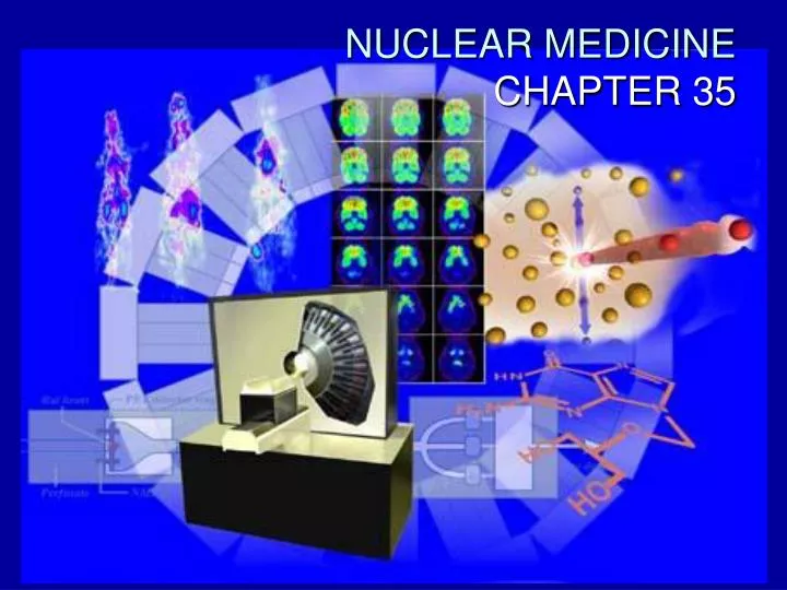
NUCLEAR MEDICINE CHAPTER 35
Topics • Nuclear Medicine licensure • Radiopharmaceuticals • Anatomy vs. Physiology • Physical principals of creating the image
Nuclear Medicine • RT (NM) • ARRT or NMTCB
NUCLEAR MEDICINE • IMAGING SPECIALITY THAT FOCUSES ON THE USE OF RADIOACTIVE MATERIALS CALL “RADIOPHARMACEUTICALS” FOR DIAGNOSIS, THERAPY, AND MEDICAL RESEARCH.
NM • DETERMINE THE CAUSE OF A MEDICAL PROBLEM BASED ON ORGAN OR TISSUE FUNCTION. • PHYSIOLOGY
Nuclear Medicine • RADIOACTIVE MATERIAL, OR “TRACER”, IS INTRODUCED INTO THE BODY BY INJECTION, SWALLOWING, OR INHALATION. • DIFFERENT TRACERS ARE USED TO STUDY DIFFERENT PARTS OF THE BODY
TRACERS • ARE SELECTED THAT LOCALIZE IN SPECIFIC ORGANS OR TISSUES • Ex: GLUCOSE • THE AMOUNT OF RADIOACTIVE TRACER MATERIAL IS SELECTED CAREFULLY TO PROVIDE THE LOWEST AMOUNT OF RADIATION EXPOSURE
RADIOACTIVE TRACERS • PRODUCE ALPHA,GAMMA or BETA EMISSION FROM WITHIN THE ORGAN BEING STUDIED • EMISSIONS ARE TRANSFORMED INTO IMAGE THAT PROVIDE INFORMATION ABOUT THE FUNCTION OF THE ORGAN OR SYSTEM BEING STUDIED.
FUNCTION VS ANATOMY • THE EMPHASIS OF NUCLEAR MEDICINE STUDIES IS MORE ON FUNCTION AND CHEMISTRY THAN ANATOMICAL STRUCTURE
HISTORY • A FEW MONTHS AFTER “WILLIE” DISCOVERED X-RAYS • HENRI BECQUEREL DISCOVERED NATURALLY OCCURRING RADIOACTIVE SUBSTANCES. • WHAT YEAR WAS THAT?
RADIOACTIVITY • IS USED TO DESCRIBE THE RADIATION OF ENERGY IN THE FORM OF HIGH-SPEED ALPHA, GAMMA OR BETA PARTICLES OR WAVES (GAMMA RAYS), FROM THE NUCLEUS OF AN ATOM
THYROID • Was one of the first organs to be examined by Nuclear Medicine studies in the 1940s-1950s • Endocrine emphasis, initially using iodine-131 to diagnose and then treat thyroid disease.
Imaging Physics Describe the • X-ray • MRI • Ultrasound • Nuclear Medicine
ISOTOPES • ELEMENTS WITH THE SAME NUMBER OF PROTONS BUT A DIFFERENT NUMBER OF _?_ ARE REFERED TO AS ISOTOPES • THE NEUTRON-TO-PROTON RATIO IN THE NUCLEUS DETERMINS THE STABILITY OF THE ATOM
ISOTOPES • At certain ratios, atoms may be unstable, a process known as spontaneous decay can occur as the atom attempts to regain stability. • Any nuclide with an atomic number greater than 83 is radioactive
ISOTOPES • ENERGY IS RELEASED IN VARIOUS WAYS DURING THIS DECAY, OR RETURN TO GROUND STATE • RADIONUCLIDES DECAY BY THE EMISSION OF ALPHA, BETA, AND GAMMA RADIATION
HALF-LIFE • DESCRIBES THE TIME IT TAKES FOR A PARTICULAR RADIONUCLIDE TO DECAY TO ONE HALF OF ITS ORIGINAL ACTIVITY • HALF-LIVES OF MOST RADIONUCLIDES USED IN NUCLEAR MEDICINE RANGE FROM SEVERAL HOURS TO SEVERAL DAYS
NUCLEAR PHARMACY • NATURALLY OCCURRING RADIONUCLIDES HAVE VERY LONG HALF-LIVES AND DELIVER HIGH ABSORBED DOSE TO THE PATIENTS • NM RADIONUCLIDES ARE MAN MADE
TECHNETIUM • IS THE MOST COMMONLY USED RADIONUCLIDE IN NM TODAY
99MTc • EXHIBITS NEARLY IDEAL CHARACTERISTICISC FOR USE IN NM • SHORT PHYSICAL HALF-LIFE OF 6.04 HOURS • PRODUCES LOW ENERGY, GAMMA PHOTONS
RADIOPHARMACEUTICALS • ARE ADMINISTERED TO PATIENTS, THEY NEED TO BE STERILE
RADIONUCLIDE IS TAGGED TO A PHARMACEUTICAL PHARMACEUTICAL CHOOSEN BASED ON THE PARTICIPATION IN THE PHYSIOLOGIC FUNCTION OF A GIVEN ORGAN RADIOPHARMACEUTICALS
RADIOPHARMACEUTICALS • Radioactive material is obtained from a manufacturer, or from an in house generator system. • "milking" the generator - sodium chloride is passed over the molybdenum-99 column, which removes the radioactive material.
RADIATION SAFETY IN NM • RADIATION PROTECTION PRACTICES DIFFER SOMEWHAT FROM THE GENERAL SAFETY RULES • RADIONUCLIDES ARE LIQUID, SOLID, OR GASEOUS FORM. • BECAUSE OF THE NATURE OF DECAY, RADIATION EMITS AFTER INJECTION
ALARA • Dosimeters • Ring dosimeters • SPILLED CHEMICALS MUST BE CLEANED UP IMMEDIATELY
INSTRUMENTATION • RADIOACTIVE DETECTORS • GEIGER COUNTER • DOSE CALIBRATOR
NM diagnostic procedure CXR UGI series Lower GI series CT Head CT Chest CT Abdomen 4.6 mSv 0.008 mSv 4.5 mSv 6 mSv 1.5 mSv 5.4 mSv 3.9 mSV – 6.1 mSv Exposure vs Effective dose the mean effective dose:
Gamma CameraSCINTILLATION DETECTORS • Used for NM image production • GIVE OFF LIGHT IN THE PRESENCE OF IONIZING RADIATION (GAMMA RAYS) • Scintillation detectors were used in the development of the first-generation NM scanner – the rectilinear scanner, 1950
Modern-day Gamma Camera • Rectilinear scanners have evolved in complex imaging systems – gamma cameras • Still use scintillation detectors: use thalliumactivated sodium iodide crystal to detect and transform radioactive emissions into light photons. Light photons are amplified and an image is recorded
Gamma Cameras • Can be stationary or mobile • Mobile cameras can move throughout the hospital or taken to SNF facilities. • Mobile cameras have limitations of smaller field of view and quality of images
Gamma rays • What is isotropic emission • Where does isotropic emission occur when producing x-rays?
Gamma Camera • Collimator: used to separate gamma rays and keep scattered rays from entering the scintillation crystal • Resolution and sensitivity are terms used to describe the physical characteristics of collimators • Made of material with a high atomic number, lead, to absorb scattered gamma rays
Crystals • Scintillation crystals commonly used are sodium iodide with trace amounts of thallim to increase light production • Thicker crystals are better for imaging radiopharmaceuticals with higher energies, but have decreased resolution • Thinner crystals provide improved resolution but not efficient with higher kV
PMTs • Photomultiplier tubes are attached to the back of the crystals • PMTs are used to detect and convert light photons emitted from the crystal into and electronic signal that amplifies the original photon signal • The electronic signal is displayed on a cathode ray tube for filming or reading
Sodium iodide • Gamma photons interact with atoms 3 ways • Photoelectric effect • Compton scattering • Pair production
Gamma Camera Systems • Standard camera: Single detector that is moved in various positions around the PT • Dual-head camera: Allows simultaneous anterior and posterior imaging and may be used for whole-body bone or tumor imaging. • Triple-head systems may be used for brain and heart studies