Download
1 / 54
570 likes | 936 Views
Overview of Hematopoietic Diseases and Their Treatments Gregory Monohan, M.D. Kentucky Cancer Registry 2012. WHO Classification of Tumors of Hematopoietic and Lymphoid Tissues. >20 Types of Leukemia >30 Types of Lymphomas CANCEROUS BLOOD CELL. Hematopoiesis. Hematopoiesis (Microenvironment).

E N D
Overview of Hematopoietic Diseases and Their TreatmentsGregory Monohan, M.D. Kentucky Cancer Registry 2012
WHO Classification of Tumors of Hematopoietic and Lymphoid Tissues • >20 Types of Leukemia • >30 Types of Lymphomas CANCEROUS BLOOD CELL
Hematopoiesis (Microenvironment) ? Proteoglycans Collagens Fibronectin Laminin Adipocyte Smooth Muscle Smooth Muscle PHSC Macrophage Tenascin Osteoclast Fibroblast Thrombospondin
CANCER BIOLOGY • THEORETICALLY: • 1/1,000,000 mistake in DNA replication made • 1/1,000,000 chance the mistake is in crucial DNA sequence that is missed by DNA repair genes. • 1 WBC created daily with significant DNA damage (potentially cancerous).
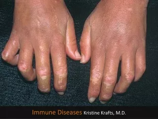
CANCER BIOLOGY • Occurs when accumulation of DNA damage leads to a cell that doesn’t die (anti-apoptotic) and can pass on cellular survival characteristics to progeny.
Maturation Arrest AML ALL NHL APL CLL HL MPDO MDS Multiple Myeloma CML
CANCER CHARACTERISTICS Cancers tend to be more metabolically active than other tissues. Higher rate of mitosis, DNA replication.
Antimetabolites (incorporate into DNA, affect folate pathways) Alkylating Agents (attack DNA) Anthracyclines (Free radicals – damage DNA) Platins (Cross-link DNA) Taxanes (bind microtubules) Vinca Alkaloids (Binds tubulin) Camptothecins (topoisomerase I inhibitors) Etoposide (topoisomerase II inhibitors) CANCER THERAPY
VEGF EGF-R C-KIT BCR-ABL TARGETED THERAPY
Backbone of Treatment Hematologic Malignancy Recovery Chemotherapy Immunotherapy • Cytopenias • Fatigue • Transfusions • Infections • Bleeding
Bone Marrow Transplant • Autologous • Recipient receives own stem cells. • Allogeniec • Recipient receives Donor derived stem cells. • Matched Related • Matched Unrelated • Mismatched • Haplo-identical • Cord Blood
Myeloid Hematologic Disease AML APL MPDO MDS CML
MyeloproliferativeNeoplasms (MPN or MPDO) • Chronic Myeloid Leukemia • Polycythaemia Vera • Primary Myelofibrosis • Essential Thrombocythaemia • Mastocytosis (Cutaneous,Systemic,Leukemic) • Chronic Neutrophilic Leukemia • Chronic Eosinophilic Leukemia • Unclassifiable
CML • 1-2/100,000 • Described by presence of BCR-ABL1 fusion gene [t(9:22)] leads to an enzyme that remains constituently active. • Bi- or Tri-phasic (Chronic phase; Accelerated phase; Blast phase) • Treatment: Tyrosine Kinase Inhibitors (Targeted therapy) treatment of choice while reserving leukemic chemotherapy +/- BMT for advanced or referactory disease.
CML Survival by Year of Presentation 1.0 TKI (92%) 0.8 0.6 1991-2000 Proportion Alive 0.4 1981-1990 0.2 1970-1980 0.0 0 2 4 6 8 10 12 14 16 18 Years from referral
AML • AML with recurrent genetic abnormalities • T(8:21); inv(16); t(15:17) [APL] • 11q23; inv(3); • Therapy related AML • AML arising from MDS • AML classified morphologically/immunophenotypically • Differentiation, wit/without maturation, myelomonocytic, monoblastic, erythroid, basophilic, megakaryoblastic.
AML Therapy • Conventional Treatment • Induction chemotherapy (1 month in hospital, 1 week of chemotherapy, then 3 weeks to recover in remission) • Consolidation chemotherapy (3-4 cycles, 6 days hospitalized, recover at home) • High Risk Treatment • As above, but would move directly into BMT when patient is in first remission, donor identified, and patient healthy.
Epidemiology • Median age 70 • 3-5/100,000 incidence • >20/100,000 in > 70 yo population • @11,000 cases / year in US
Aul et al (Germany) Radlund et al (Sweden) 25 20 15 Incidence/100,000 population/yr 10 5 0 <50 yr 5069 yr >70 yr All ages MDS: Incidence Aul C et al. Leuk Res. 1998;22:93 Radlund A et al. Eur J Haematol. 1995;54:153
Clinical Manefestations • Majority of symptoms secondary to cytopenias: • Anemia • Infections • Transfusion dependence • Patechaeie, bruising • Infrequent organomegally
Dysgranulopoiesis Dyserythropoeisis Dyspoetic Megakaryocytes
International Prognostic Scoring System (IPSS) All 3 prognostic variables required to generate IPSS score Score Value Prognostic variable 0 0.5 1.0 1.5 2.0 Bone marrow blasts (%) <5 5–10 – 11–19 20–30 Karyotype* Good Intermediate Poor – Number of cytopenias** 0/1 2/3 – – – *Good=normal, -Y, del(5q), del(20q); Intermediate=other karyotypic abnormalities; Poor=complex (3 abnormalities) or chromosome 7 abnormalities **Hgb <10 g/dL; ANC <1800/L; platelet count <100,000/L Survival and Evolution to AML by IPSS Parameter Low Int-1 Int-2 High Score 0 0.5–1.0 1.5–2.0 2.5 Median survival, yr 5.7 3.5 1.2 0.4 25% AML evolution, median, yr 9.4 3.3 1.1 0.2 Greenberg P et al. Blood. 1997;89:2079
100 100 90 90 80 80 70 70 60 60 50 50 40 40 30 30 20 20 10 10 0 0 13 13 15 14 18 10 11 14 15 12 18 12 11 10 16 16 17 17 1 9 8 7 6 4 2 1 3 2 3 4 6 7 8 9 0 0 5 5 Survival and AML Evolution by IPSS Classification Survival AML Evolution Low 235 pts Int-1 295 pts Int-2 171 pts High 58 pts Low 267 pts Int-1 314 pts Int-2 179 pts High 56 pts Percent Percent Years Years From diagnosis in untreated patients Reprinted with permission from Greenberg P et al. Blood. 1997;89:2079
Lymphoid Hematologic Disease ALL NHL CLL HL Multiple Myeloma
CLL • 2-6/100,000 • Clinical Presentation • Asymptomatic • Splenomegaly, Lymphadenopathy • Infections, Bleeding, Hemolytic Anemia • Treatment • Observation • Chemotherapy • BMT for aggressive or refractory Disease
ALL • 75% cases are in children < 6 year old. • 1-4.75/100,000 • In children, prolonged alternating chemotherapy (Induction, Consolidation, Intensification, and Maintenance) leads to >80% cure rate. • In adults, <50% cure rates and healthy patients are selected for BMT
NHL • Indolent • Follicular NHL • Aggressive • Diffuse Large Cell NHL • Mantle Cell Lymphoma • Burkitt Lymphoma
Follicular NHL • Clinically • Asymptomatic • Lymphadenopathy • Splenomegaly • Treatment • Observation until symptomatic • Chemotherapy • Immunotherapy • BMT for refractory or aggressive disease
DLBCL • Aggressive, rapidly enlarging tumor mass • Nodal or extranodal • @50% are stage 1 or 2. • Respond very well to chemotherapy +/- radiation therapy, >50% cure even in stage 4 disease. • In relapse, many patients can be salvaged with Autologous BMT.
Hodgkin’s Lymphoma • Classical Hodgkin’s Lymphoma • Nodular sclerosis • Mixed Cellularity • Lymphocyte Rich • Lymphocyte Depleted • Nodular Lymphocyte Predominate HL
Hodgkin’s Lymphoma • Usually present with Lymphadenopathy. • >85% curable with Chemotherapy and/or Radiation therapy. • Salvage relapsed patients with Autologous BMT.
International Myeloma Working Group Criteria • M-protein in serum and/or urine • Marrow plasma cells (clonal) or plasmacytoma • Related Organ or Tissue Impairment: • Calcium elevated. • Renal insufficiency • Anemia (Hgb <10 or 2mg/dL below LLN • Bone lytic lesions or osteoporosis with compression fracture. • Other: hyperviscosity, amyloidosis, recurrent bacterial infections. British Journal of Haematology 2003;121:749-757
PLASMA CELLS Normal Myeloma
Disease Manifestations of MM Bone Destruction Bone Pain Cytokine Release Hypercalcemia Anemia Marrow Infiltration Multiple Myeloma Immune Deficiency Monoclonal Protein Light-Chain Deposition Disease Infection Renal Failure Clotting abnormalities Hyperviscosity Amyloidosis