Download
1 / 18
200 likes | 371 Views
PHACOMATOSES. 1. Neurofibromatosis. Type I (NF-1) - von Recklinghausen disease Type II (NF-2) - bilateral acoustic neuromas. 2. Tuberous sclerosis (Bourneville disease). 3. von-Hippel-Lindau syndrome. 4. Sturge-Weber syndrome. Neurofibromatosis type-1 - (NF-1).

E N D
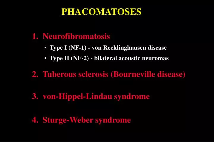
PHACOMATOSES 1. Neurofibromatosis • Type I (NF-1) - von Recklinghausen disease • Type II (NF-2) - bilateral acoustic neuromas 2. Tuberous sclerosis (Bourneville disease) 3. von-Hippel-Lindau syndrome 4. Sturge-Weber syndrome
Neurofibromatosis type-1 - (NF-1) • Most common phacomatosis • Affects 1:4000 individuals • Presents in childhood • Gene localized to chromosome 17q11 Café-au-lait spots Increase in size and number throughout childhood Appear during first year of life
Fibroma molluscum in NF-1 • Appear at puberty • Pedunculated, flabby nodulesconsisting of • neurofibromas or schwannomas • Increase in number • throughout life • Frequently widely distributed
Plexiform neurofibroma in NF-1 • Appear during childhood • Large and ill-defined • May be associated with • overgrowth of overlying skin
Skeletal defects in NF-1 • Facial hemiatrophy • Mild head enlargement - uncommon • Other - scoliosis, short stature, thinning of • long bones
Orbital lesions in NF-1 Optic nerve glioma in about 15% Spheno-orbital encephalocele • Sagittal MRI scan of optic nerve glioma • invading hypothalamus • Glioma may be unilateral or bilateral • Axial CT scan of congenital absence of • left greater wing of sphenoid bone • Causes pulsating proptosis without bruit
Eyelid neurofibromas in NF-1 Nodular Plexiform May cause mechanical ptosis May be associated with glaucoma
Intraocular lesions in NF-1 Lisch nodules Congenital ectropion uveae Very common - eventually present in 95% of cases Uncommon - may be associated with glaucoma Choroidal naevi Retinal astrocytomas Rare - identical to those seen in tuberous sclerosis Common - may be multifocal and bilateral
Very common - presenile cataract Ocular features of NF-2 Common - combined hamartomas of RPE and retina
Tuberous sclerosis (Bournevill disease) • Autosomal dominant • Triad - mental handicap, epilepsy, adenoma sebaceum Adenoma sebaceum Ash leaf spots Shagreen patches • Around nose and • cheeks • Appear after age 1 • and slowly enlarge • Hypopigmented skin patches • In infants best detected using • ultraviolet light (Wood’s lamp) • Diffuse thickening over • lumbar region • Present in 40%
Systemic hamartomas in tuberous sclerosis Astrocytic cerebral hamartomas Visceral and subungual hamartomas • Usually asymptomatic and • innocuous • Kidneys (angiomyolipoma), heart • (rhabdomyoma) • Slow-growing periventricular tumours • May cause hydrocephalus, epilepsy and • mental retardation
Retinal astrocytomas in tuberous scleritis • Innocuous tumour present in 50% of patients • May be multiple and bilateral Early Semitranslucent nodule White plaque Advanced Dense white tumour Mulberry-like tumour
Systemic features of v-H-L syndrome Autosomal dominant CNS Haemangioblastoma Visceral tumours MRI of spinal cord tumour • Tumours - renal • carcinoma and • phaeochromocytoma • Cysts - kidneys, liver, • pancreas, epididymis, • ovary and lungs • Polycythaemia Angiogram of cerebellar tumour
Retinal capillary haemangioma in v-H-L syndrome • Vision-threatening tumour present in 50% of patients • May be multiple and bilateral Early Tiny lesion between arteriole and venuole Small red nodule Advanced Associated dilatation and tortuosity of feeder vessels Round orange-red mass
Complications of retinal capillary haemangioma Leakage Exudative retinal detachment Hard exudate formation Epiretinal membrane formation
Treatment options of retinal capillary haemangioma • Argon laser photocoagulation - small peripheral tumours • Cryotherapy - larger peripheral tumours • External beam radiotherapy - if unresponsive to cryotherapy Before treatment - dilated feeder vessels Following treatment - normal feeder vessels
Systemic features of Sturge-Weber syndrome Naevus flammeus Meningeal haemangioma • Congenital, does not blanche • with pressure • Associated with ipsilateral • glaucoma in 30% of cases • CT scan showing left • parietal haemangioma • Complications - mental handicap, • epilepsy and hemiparesis
Ocular features of Sturge-Weber syndrome Glaucoma Buphthalmos in 60% May be associated with episcleral haemangioma Diffuse choroidal haemangioma Affected eye Normal eye