Download
1 / 47
520 likes | 920 Views
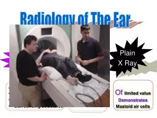
Radiology of the abdomen. Radiological modalities. X – Ray Flouroscopy U/S CT scan MRI. X - Ray. It is ionizing radiation – radiation hazard. It is useful in assessing the bones, bowel gases (obstruction) and calcification. Normal AXR. Normal AXR. Gas in stomach. Liver.

E N D
Radiological modalities • X – Ray • Flouroscopy • U/S • CT scan • MRI
X - Ray • It is ionizing radiation – radiation hazard. • It is useful in assessing the bones, bowel gases (obstruction) and calcification.
Normal AXR Gas in stomach Liver Splenic flexure T12 11th rib Psoas margin Left kidney Hepatic flexure Transverse colon Iliac crest Gas in sigmoid Sacrum Gas in caecum SI joint Bladder Femoral head
Gas pattern • Stomach • Almost always air in stomach • Small bowel • Usually small amount of air in 2 or 3 loops • Large bowel • Almost always air in rectum and sigmoid • Varying amount of gas in rest of large bowel What is normal?
3, 6, 9 RULE Maximum Normal Diameter of bowel Small bowel 3cm Large bowel 6cm Caecum 9cm
Mechanical SBO • Dilated small bowel • Fighting loops (visible loops, lying transversely, with air-fluid levels at different levels) • Little gas in colon, especially rectum
SBO Erect SBO Supine Air fluid levels
Step ladder appearance • Loops arrange themselves from left upper to right lower quadrant in distal SBO
Double Bubble Sign Duodenal Atresia
Mechanical LBO • Colon dilates from point of obstruction backwards • Little/no air fluid levels (colon reabsorbs water) • Little or no air in rectum/sigmoid
Coffee Bean SignSigmoid volvulus Massively dilated sigmoid loop
Thumbprinting The distance between loops of bowel is increased due to thickening of the bowel wall. The haustral folds are very thick, leading to a sign known as 'thumbprinting.'
Extraluminal air • TYPES • Pneumoperitoneum/free air/intraperitoneal air • Retroperintonealair • Air in the bowel wall (pneumatosisintestinalis) • Air in the biliary system (pneumobilia)
Upright film best • The patient should be positioned sitting upright for 10-20 minutes prior to acquiring the erect chest X-ray image. • This allows any free intra-abdominal gas to rise up, forming a crescent beneath the diaphragm. It is said that as little as 1ml of gas can be detected in this way.
Free AirCauses • Rupture of a hollow viscus • Perforated peptic ulcer • Trauma • Perforated diverticulitis (usually seals off) • Perforated carcinoma • Post-op 5-7 days normal, should get less with successive studies *NOT ruptured appendix (seals off)
Signs of free air • Crescent sign • Riglers sign • Football sign • Falciform ligament sign
Crescent Sign II Free air under the diaphragm Best demonstrated on upright chest x rays or left latdecub Easier to see under right diaphragm ? Why?
Rigler’s Sign Bowel wall visualised on both sides due to intra and extraluminal air Usually large amounts of free air May be confused with overlapping loops of bowel, confirm with upright view
Football Sign Seen with massive pneumoperitoneum Most often in children with necrotisingenterocolitis In supine position air collects anterior to abdominal viscera Paediatric Adult
Falciform ligament sign Normally invisible. Supine film, free air rises over anterior surface of liver
Soft tissue masses • Organomegaly • Know normal landmarks CT, US and MRI have essentially replaced conventional radiography in the assessment of organomegaly and soft tissue masses
Abdominal Calcifications Location Pattern
Calcified fibroids Calcified enteric lymph nodes Calcified pancreas Floccular
Bladder calculi Lamellar
Renal calculi Pelvicalyceal calcifications
StaghornCalcification Renal stones are often small, but if large can fill the renal pelvis or a calyx, taking on its shape which is likened to a staghorn. Tubular
Renal calculi Parenchymal calcification Nephrocalcinosis Uncommonly the renal parenchyma can become calcified. This is known as nephrocalcinosis, a condition found in disease entities such as medullary sponge kidney or hyperparathyroidism. Flocculent
Floruscopy • We are using a contrast material for better visualization of hollow organs, such as bowel loops and KUB. • It is useful to assess the mucosal pathology. • We can use either oral or rectal contrast • If we use rectal contrast; we can use either: • Single contrast barium enema. • Double contrast barium enema.
4 What type of this study? Single or double? 5 6 3 7 2 8 1
4 5 6 Rectum Sigmoid colon Descending colon Splenic flexure Transverse colon Hepatic flexure Ascending colon cecum 3 7 2 8 1
Abnormal study Colon Cancer (apple core sign)
Lead pipe colon • Shortening of colon secondary to fibrosis • Loss of haustration • Ulcerative colitis
Abnormal Normal
CT Scan • It is an ionizing radiation. • Corss-sectional imaging. • Better anatomical visualization.
5 2 1 6 4 3
5 2 1 6 4 3 1- Rectum 2-Sigmoid colon 3-Descending colon 4-Ascending colon 5-Transverse colon 6-Cecum
3 2 4 1 5 6
3 2 4 1 5 6 Descending colon Splenic flexure Hepatic flexure Ascending colon cecum Sigmoid colon