Download
1 / 32
400 likes | 744 Views
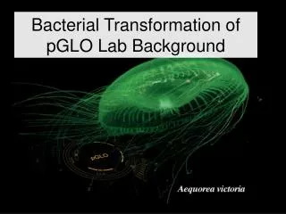
pGLO™ Transformation and Purification of Green Fluorescent Protein (GFP). Discovery of GFP. Naturally produced in Jellyfish– Aequorea victoria Discovered in 1960 ’ s. Osamu Shimomura first isolated GFP from the jellyfish Aequorea victoria,

E N D
pGLO™ Transformation and Purification of Green Fluorescent Protein (GFP)
Discovery of GFP • Naturally produced in Jellyfish– Aequorea victoria • Discovered in 1960’s
Osamu Shimomura first isolated GFP from the jellyfish Aequorea victoria, Martin Chalfie demonstrated the value of GFP as a luminous genetic tag for various biological phenomena. Roger Y. Tsien contributed to our general understanding of how GFP fluoresces. He also extended the colour palette beyond green allowing researchers to give various proteins and cells different colours. Osamu Shimomura Martin Chalfie Roger Y. Tsien
Osamu Shimomura: first isolated GFP from the jellyfish Aequorea victoria,
Martin Chalfie: C. elegans glowing with green fluorescent protein
Roger Tsien: range of fluorescent proteins have brought some colour to laboratory work.
The barrel structure of GFP The tertiary structure of GFP is barrel-like, consisting of 11 beta sheets depicted as the green ribbons and an internal chromophore of three adjacent amino acids, depicted as green spheres
Cyclization of the tripeptide Ser-Tyr-Gly: The active chromophore of GFP is comprised of three adjacent amino acids in the primary amino acid chain. The three amino acids are enzymatically converted to an active cyclic chromophore in vivo
Using GFP as a biological tracer http://www.conncoll.edu/ccacad/zimmer/GFP-ww/prasher.html With permission from Marc Zimmer
Links to Real-world • GFP is a visual marker • Study of biological processes (example: synthesis of proteins) • Localization and regulation of gene expression • Cell movement • Cell fate during development • Formation of different organs • Screenable marker to identify transgenic organisms
What is a plasmid? • A circular piece of autonomously replicating DNA • Originally evolved by bacteria • May express antibiotic resistance gene • or be modified to express proteins of interest
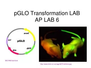
Bacterial Transformation Cell wall GFP Bacterial chromosomal DNA Beta lactamase (ampicillin resistance) pGLO plasmids
GeneExpression • Beta Lactamase • Ampicillin resistance • Green Fluorescent Protein (GFP) • Aequorea victoria jellyfish gene • araC regulator protein • Regulates GFP transcription
ara Operon lac Operon araC B A D LacI Z Y A Effector (Arabinose) Effector(Lactose) araC B A D LacI Z Y A RNA Polymerase RNA Polymerase B A D araC Z Y A Transcriptional Regulation
ara GFP Operon ara Operon araC GFP Gene araC B A D Effector(Arabinose) Effector (Arabinose) araC B A D araC GFP Gene RNA Polymerase RNA Polymerase B A D araC araC GFP Gene Gene Regulation
LB/Amp LB/Amp/Ara LB Now, prepare 3 kinds of plates 1. LB 2. LB + Amp+ (3 plates) 3. LB + Amp+ + Arabinose (2 plates)
1: LB plates 2、3、4: LB +Amp+plates 5、6、7: LB+ Amp++Arabinose plates
LB/Amp LB/Amp/Ara Store concentration Working concentration 10 mg/ml 50mg/ml Amp+ Arabinose 200mg/ml 50mg/ml 50ml LB medium+250ml Amp+ 50ml LB medium+250ml Amp++12.5ml Arabinose Ampicillin will be degraded in high temperature!
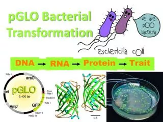
DNA RNA Protein Trait Central Framework of Molecular Biology
What is Transformation? Uptake of foreign DNA, often a circular plasmid GFP Beta-lactamase Ampicillin Resistance
Methods of Transformation Electroporation Electrical shock makes cell membranes permeable to DNA Calcium Chloride/Heat-Shock Chemically-competent cells uptake DNA after heat shock
Reasons for Performing Each Transformation Step? Transformation solution = CaCI2 Positive charge of Ca++ ions shields negative charge of DNA phosphates Ca++ O Ca++ O P O Base O O CH2 Sugar O Ca++ O O P Base O O CH2 Sugar OH
Why Perform Each Transformation Step? Cell wall GFP 2. Incubate on ice slows fluid cell membrane 3. Heat-shock Increases permeability of membranes 4. Nutrient broth incubation Allows beta-lactamase expression Beta-lactamase (ampicillin resistance)
Transformation Procedure Prepare 2 tubes, add 250ml of 50mMCacl2 Suspend 2-4 bacterial colonies in Cacl2 add 1ml of 0.2mg/ml pGLO plasmid DNA to one tube, the other one leave as control Extra competent cells: Add 1ml of pGLO plasmid • Place tubes on ice(10mins) • Heat-shock at 42°C for 50sec~1min and place on ice(1min) • Add 250ml LB medium and Incubate at 37oC for10 mins • Centrifuge ,get rid of 300-350ml supernatant, re-suspend, • Streak plates • Incubate LB platesin incubator o/n
Transformation Procedure Overview Day 2 Day 1
Make sure to streak to correct plates! extra competent cells
LB/Amp LB/Amp/Ara LB Grow?Glow? • Follow protocol • On which plates will colonies grow? • Which colonies will glow?