Download
1 / 93
930 likes | 1.12k Views
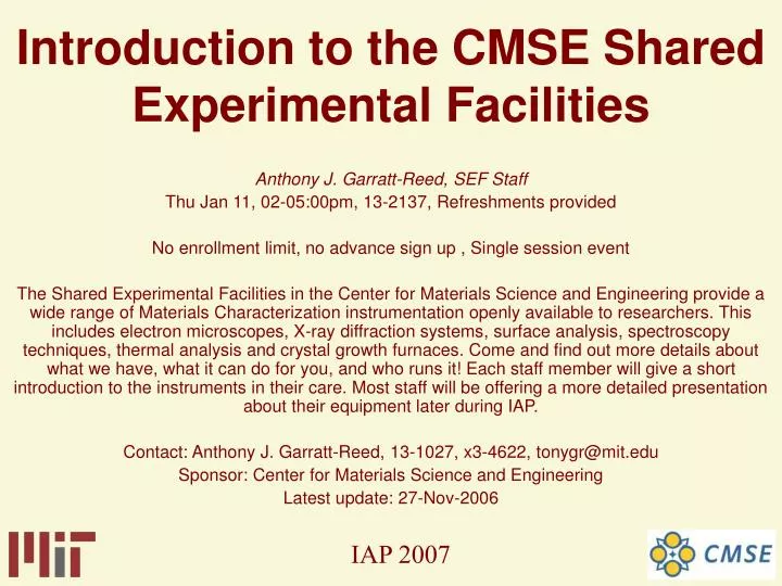
Introduction to the CMSE Shared Experimental Facilities. Anthony J. Garratt-Reed, SEF Staff Thu Jan 11, 02-05:00pm, 13-2137, Refreshments provided No enrollment limit, no advance sign up , Single session event

E N D
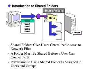
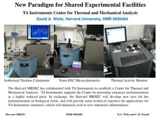
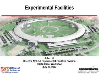
Introduction to the CMSE Shared Experimental Facilities Anthony J. Garratt-Reed, SEF Staff Thu Jan 11, 02-05:00pm, 13-2137, Refreshments provided No enrollment limit, no advance sign up , Single session event The Shared Experimental Facilities in the Center for Materials Science and Engineering provide a wide range of Materials Characterization instrumentation openly available to researchers. This includes electron microscopes, X-ray diffraction systems, surface analysis, spectroscopy techniques, thermal analysis and crystal growth furnaces. Come and find out more details about what we have, what it can do for you, and who runs it! Each staff member will give a short introduction to the instruments in their care. Most staff will be offering a more detailed presentation about their equipment later during IAP. Contact: Anthony J. Garratt-Reed, 13-1027, x3-4622, tonygr@mit.edu Sponsor: Center for Materials Science and Engineering Latest update: 27-Nov-2006
Tim McClure Analysis Shared Experimental Facility Project Technician Office: 13-4149 Phone: (617) 258-6470 Email: mtim@mit.edu http://mit.edu/mtim/www MIT Center for Material Science and Engineering 77 Massachusetts Avenue, Cambridge, Massachusetts 02139
Analysis SEF Staff • Dr. Shaoyan Chu sc79@mit.edu X3-0054 • Tim McClure mtim@mit.edu X8-6470 • Elisabeth Shaw elshaw@mit.edu X3-5045
13-4111 13-4139 13-4147 13-4137 13-4149 Analysis SEF
Analysis SEF • Thermal Analysis 13-4111 • Dr. Shaoyan Chu sc79@mit.edu • Surface Analysis 13-4137 • Libby Shaw elshaw@mit.edu • Spectroscopy 13-4139 • Tim McClure mtim@mit.edu • AFM/Thin Film 13-4147 • Libby Shaw elshaw@mit.edu
Instrumentation in 13-4139 Spectroscopy Microscopy Thin Film Support
Instrumentation in 13-4139 Spectroscopy FTIR, Raman, UV/VIS/NIR, Fluorimeter Microscopy Thin Film Support
Instrumentation in 13-4139 Spectroscopy FTIR, Raman, UV/VIS/NIR, Fluorimeter Microscopy Optical, Thermal, FTIR, Raman Thin Film Support
Instrumentation in 13-4139 Spectroscopy FTIR, Raman, UV/VIS/NIR, Fluorimeter Microscopy Optical, Thermal, FTIR, Raman Thin Film Profilometer, Flexus, Thermal Co-Evaporator, Plasma Cleaner Support
Instrumentation in 13-4139 Spectroscopy FTIR, Raman, UV/VIS/NIR, Fluorimeter Microscopy Optical, Thermal, FTIR, Raman Thin Film Profilometer, Flexus, Thermal Co-Evaporator, Plasma Cleaner Support Temperature Controlled Microscope Stages, Cryostat, Lasers, Exhaust Hood, Oven
Spectroscopy There are many types of spectroscopic analysis techniques XRAY XPS ICP Etc.
Optical Spectroscopy Methods Absorption Emisson Luminescence Scattering
Absorption Spectroscopy • Absorbance or the ratio of transmitted to incident radiant power • Atomic absorption • UV/VIS molecular absorption • IR absorption
Emission Spectroscopy • Radiant power of emission • ICP • Spark • Flame • DC Arc
Luminescence Spectroscopy • Radiant power of luminescence • Molecular Fluorescence • Phosphorescence • Chemical and Bioluminescence • Atomic Fluorescence
Scattering Spectroscopy • Radiant power of scattering • Raman Scattering • Mie scattering • turbidity
Optical Spectroscopy Interaction of optical electromagnetic radiation with matter Vibrational
Spectra Intramolecular Vibrations
Stretching Vibrations • In a simple diatomic molecule A-B the only vibration which can occur is a periodic stretching along the A-B bond. • Stretching vibrations resemble the oscillations of two bodies connected by a spring.
Hooke’s Law For stretching of the bond A-B, the vibrational frequency v (cm-1) is given by the equation: v = (1/2c) (f/)1/2 c = Velocity of light f = Force constant of the bond = reduced mass of the system
Reduced Mass Equation = (mA.mB)/(mA+mB) = reduced mass of the system mA and mB are the individual masses of A and B
Spectrochemical Analysis Is used to monitor • Water we drink • Food we eat • Status of human health • Quality of the environment
Spectroscopic Intruments • Fourier Transform Infrared (FTIR) -Thermo Nicolet • Magna 860 Bench • Nic Plan Microscope • Raman Microprobe -Kaiser • Hololab 5000R Modular Research System • Argon Ion (514.5nm), Titanium Sapphire (785nm) CW Lasers, Diode (785nm) • UV/Vis Spectrometers -Cary • 5e • 500i • Fluorimeter -PTI • QM6/2006
Infrared Spectra The absorption or emission spectrum arising from the rotational and vibrational motions of a molecule which is not electronically excited is mostly in the infrared region.
IR Active Vibration Selection Rule There must be a net change in permanent dipole moment during the vibration
Vibrational Energies • The energy of a vibrational mode depends on • molecular structure • and environment. • Atomic mass • bond order • molecular substituents • molecular geometry • hydrogen bonding • all effect the vibrational force constant
IR Spectroscopy Studies Identification of a Substance Determination of Molecular structure Determination of Purity Reaction Kinetic Studies Fundamental Studies of Molecules
Spectra • A unique physical property and is characteristic of the molecule • The infrared spectrum can be used as a fingerprint for identification • Compare with previously recorded reference spectra
Nicolet Magna 860 FTIR Spectrometer • Beam splitter, Detector, Source combinations allow extended range measurement in the Mid, Near and Far IR ranges. • Accessories • Emission Experiments • Horizontal Attenuated Total Reflectance • Variable Angle Reflectance • Optical Cryostat • Solid, Liquid, Gas Sampling Accessories
Spectra Tech Nic Plan FTIR Microscope • This Microscope works with the Nicolet Magna 860 Spectrometer. • Spot sizes as small as 10 microns can be sampled. • It has a motorized stage with Sample Mapping and Video Capture capabilities. • Accessories • ATR Objective • 15X, 32X IR Objectives • Micro Compression Cell • View Thru Aperture
Raman Effect • Classical • Perturbation of the molecule’s electric field • Quantum mechanical • scattering is an excitation to a virtual state • lower in energy than a real electronic transition with nearly coincident de-excitation and a change in vibrational energy • The scattering event occurs in 10-14 seconds or less
Raman Spectroscopy • The Raman effect arises when a photon is incident on a molecule and interacts with the electric dipole of the molecule. • It is a form of electronic spectroscopy • Vibronic is more accurate
Raman Spectrum The difference in energy between the incident photon and the Raman scattered photon is equal to the energy of a vibration of the scattering molecule. A plot of intensity of scattered light versus energy difference is a Raman spectrum
Weak Raman Scatterers Highly polar moiety vibrations O-H bond An external electric field can not induce a large change in the dipole moment Stretching or bending the bond does not change this
Strong Raman Scatterers • Moieties with distributed electron clouds • carbon-carbon double bonds • The electron cloud of the double bond is easily distorted in an external electric field • Bending or stretching the bond changes the distribution of electron density substantially, and causes a large change in induced dipole moment
IR & Raman Molecular Frequencies Infrared absorption frequencies frequently agree with the frequency shifts found in the Raman effect Not always true Depends on the symmetry of the molecule
Raman Uses Vibrational Raman spectroscopy is not limited to intramolecular vibrations Crystal lattice vibrations and other motions of extended solids are Raman-active Polymers Semiconductors
Vibration/Rotation Spectra • Gas phase • rotational structure is resolvable on vibrational transitions • The resulting vibration/rotation spectra are widely used to study combustion and gas phase reactions
Vibrations in Solids Spectra of crystals are almost always sharper than those of melts or solutions. Spectra of solids when compared to those of the solute in solution or their melts are invariably much more complex. The frequencies of the bands in the solid are shifted from those of itself in the liquid phase. Some bands can move considerably (30cm-1 is not unusual). (IJVS)
Raman Microprobe • Kaiser Optical Systems • Modular 5000R Research Grade Raman • Leica Microscope • Prior Motorized Stage • 514.5nm or 785nm Excitation Lasers • Holographic Grating Spectrometer • Andor Deep Depletion CCD 1024 X 512 • Small or Large Fibers