Download
1 / 58
580 likes | 727 Views
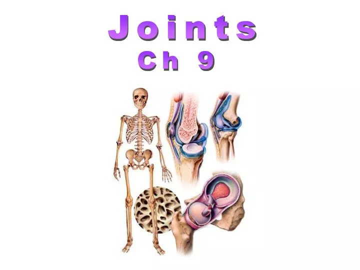
Joints. Ch 9. Joints. Articulations: The site where 2 or more bones meet. Joints are the weakest part of the skeleton. Classification Functional: Amount of movement allowed 1). Synarthroses: Immovable joints 2). Amphiarthrosis: Slightly movable joint

E N D
Joints Ch 9
Joints • Articulations: The site where 2 or more bones meet. • Joints are the weakest part of the skeleton. • Classification • Functional: Amount of movement allowed • 1). Synarthroses: Immovable joints • 2). Amphiarthrosis: Slightly movable joint • 3). Diarthroses: Fully movable joints
(b) Syndesmosis Joint held together by a ligament. Fibrous tissue can vary in length, but is longer than in sutures. Fibula Tibia Ligament Joints • Classification • Structural: based on material binding the bone. • 1). Fibrous: Bone ends united by collagenic fibers • a). Sutures • b). Syndesmoses • c). Gomphoses
Joints • Classification • 2). Cartilaginous Joints • Bones are united by cartilage • a). Synchondrosis • b). Symphyses • c). Synovial Joints
suture Fibrous Joints Immovable Joints (synarthrosis) • Bones united by ligament
Fibrous Joints Immovable Joints (synarthrosis) (syndesmosis) • Bones united by ligament
Fibrous Joints Immovable Joints (synarthrosis) Interosseous membrane (syndesmosis) • Bones united by ligament
Gomphosis enamel dentin pulp gum Socket of alveolar process root of tooth Peridontal ligament (membrane) • Ligaments hold tooth in bony socket • Immovable joint
Cartilagenous Joints Slightly Movable (ampharthrosis) and Immovable (synarthrosis) Joints • Lacks a synovial cavity • Bones connected by fibrocartilage or hyaline cartilage • 2 types • synchondrosis • symphyses
Cartilagenous Joints Immovable Joint (synchondrosis)
Cartilagenous Joints Slightly Movable Joint (ampharthrosis) pubic symphysis symphysis
pelvis ligaments femur Synovial Joints (diarthrosis)- freely moveable
hyaline cartilage synovial cavity femur Synovial Joints
Synovial Joints Shoulder joint
Knee Joint Cruciate Ligaments Fig. 9.13 The cruciate ligaments prevent undesirable movements at the knee joint. a) when the knee is flexed or extended, the anterior cruciate prevents anterior slipping movements of the tibia
Temporomandibular Joint • Complex Joint • Articular disc • Gliding above disc • Hinge below disc • Movements: • depression • elevation • protraction • retraction
Types of Synovial Joints • Planar Joint • Hinge Joint • Pivot Joint • Saddle Joint • Ball & Socket Joint • Condyloid or Ellipsoid Joint
Hinge Joint • Convex surface of bone fits in concave surface of 2nd bone • Unixlateral like a door hinge • Examples: • Knee, elbow, ankle, interphalangeal joints • Movements produced: • flexion • extension • hyperextension
Planar Joint • Bone surfaces are slightly curved • Side to side movement only • Rotation prevented by ligaments • Examples: • intercarpal to intertarsal joints • sternoclavicular joint • vertebrocostal joints
Pivot Joint • Rounded surface of bone articulates with the ring formed by the 2nd bone & ligament • Monoaxial since it only allows rotation around longitudinal axis • Examples: • proximal radioulnar joint • supination • pronation • atlanto-axial joint • Turning head side to side “no”
Saddle Joint • One bone saddle-shaped, other bone fits like a person riding on the saddle • Biaxial • circumduction allows the tip of the thumb to travel in a circle • Opposition allows thumb to touch tip of other fingers • Examples: • Trapezium of carpus and metacarple of thumb
Ball & Socket Joint • Ball fitting into a cup-like depression • Multiaxial • flexion/extension • abduction/adduction • rotation • Examples: • shoulder joint • hip joint
Condyloid Joint • Oval-shaped depression fits into oval depression • Biaxial= flex/extend or adduct/abduct is possible • Examples: • Wrist and metacarpophelangeal joints for 2 to 5 digits
Gliding (a) Gliding movements at the wrist Figure 8.5a Movements allowed by synovial joints.
Hyperextension Extension Flexion (b) Angular movements: flexion, extension, and hyperextension of the neck Figure 8.5b Movements allowed by synovial joints.
Extension Hyperextension Flexion (c) Angular movements: flexion, extension, andhyperextension of the vertebral column Figure 8.5c Movements allowed by synovial joints.
Flexion Extension Flexion Extension (d) Angular movements: flexion and extension at theshoulder and knee Figure 8.5d Movements allowed by synovial joints.
Abduction Circumduction Adduction (e) Angular movements: abduction, adduction, andcircumduction of the upper limb at the shoulder Figure 8.5e Movements allowed by synovial joints.
Rotation Lateral rotation Medial rotation (f) Rotation of the head, neck, and lower limb Figure 8.5f Movements allowed by synovial joints.
Pronation (radius rotates over ulna) Supination (radius and ulna are parallel) (a) Pronation (P) and supination (S) Figure 8.6a Special body movements.
Dorsiflexion Plantar flexion (b) Dorsiflexion and plantar flexion Figure 8.6b Special body movements.
Inversion Eversion (c) Inversion and eversion Figure 8.6c Special body movements.
Protraction of mandible Retraction of mandible (d) Protraction and retraction Figure 8.6d Special body movements.
Depression of mandible Elevation of mandible (e) Elevation and depression Figure 8.6e Special body movements.
Opposition (f) Opposition Figure 8.6f Special body movements.
Type of joint movement: • Flexion- bent knee • Extension- extend knee • Hyperextension- bring leg back • Dorsi flexion- heal • Plantar flexion- toe • Abduction- leg out • Adduction-leg in • Rotation- twisting • Circumduction- circular motion • Supination- palm up • Pronation- palm down • Eversion- foot out • Inversion- foot in • Protraction- chin forward • Retraction- chin back • Elevation- shoulders up • Depression- shoulders down
Factors Influencing Joint Stability A)The shape of articular surfaces. B) Ligaments C) Muscle Tone
Disorders and Imbalances • Bursitis • Tendonitis • Lyme disease • Ankle sprains and fractures • Osteoarthritis • Gouty Arthritis • Rheumatoid Arthritis
Disorders and Imbalances • Lyme disease
Disorders and Imbalances • Ankle Sprain Type 1
Disorders and Imbalances • Ankle Sprain Type 2
Disorders and Imbalances • Ankle Sprain Type 3
Osteoarthritis • Degenerative joint disease • aging, wear & tear • Non inflammatory • Only cartilage is affected, not synovial membrane • Deterioration of cartilage produces bone spurs • Restricts movement • Pain upon awakening—disappears with movement
Gouty Arthritis • Uric crystals build up in joints—pain • waste products of DNA & RNA metabolism • builds up in blood • deposited in cartilage causing inflammation and swelling • Bones fuse • Middle-aged men with abnormal gene