Download
1 / 3

E N D
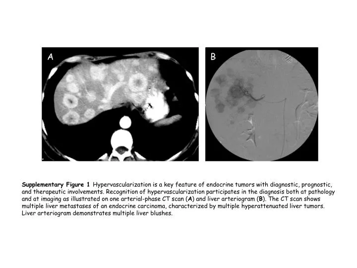
A B Supplementary Figure 1 Hypervascularization is a key feature of endocrine tumors with diagnostic, prognostic, and therapeutic involvements. Recognition of hypervascularization participates in the diagnosis both at pathology and at imaging as illustrated on one arterial-phase CT scan (A) and liver arteriogram (B). The CT scan shows multiple liver metastases of an endocrine carcinoma, characterized by multiple hyperattenuated liver tumors. Liver arteriogram demonstrates multiple liver blushes.
B A Supplementary Figure 2: Endocrine tumors represent a spectrum of tumors characterized by various ratios of differentiation to proliferation. This feature is illustrated, by somatostatin receptor scintigraphy (SRS) and PET-fluorodeoxyglucose (FDG), in two patients presenting with a metastatic endocrine carcinoma. In each case, both the static planar anterior SRS and the anterior whole-body PET-FDG are provided. Shown is a patient with a well-differentiated pancreatic endocrine carcinoma: (A) SRS depicts multiple liver foci; (B) PET-FDG was negative, suggestive of a high differentiation-to-proliferation ratio.
D C Supplementary Figure 2: panels C and D show a case of a patient with a poorly differentiated endocrine carcinoma: (C) SRS was found weakly positive depicting a minority of foci within the liver; (D) in contrast PET-FDG showed a higher number of highly intense foci within the thorax, bone and abdomen suggestive of a low differentiation over proliferation ratio.