Download
1 / 66
680 likes | 973 Views
Chapter 45. Skeletal, Muscular, and Integumentary System. Intercellular Junctions . A tight junction completely encircles an epithelial cell near its apex (top) and joins it tightly to the neighboring cells. Like a six pack, each cell have complementary groves and ridges to make it zipper like.

E N D
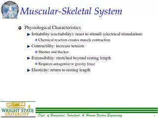
Chapter 45 Skeletal, Muscular, and Integumentary System
A tight junction completely encircles an epithelial cell near its apex (top) and joins it tightly to the neighboring cells. Like a six pack, each cell have complementary groves and ridges to make it zipper like. For nurtrient regulation it can be “unzipped” in like the small intestine
A desmosomes is like a snap on a pair of jeans. It is a patch that holds cells together and enables a tissue to resist mechanical stress, but does not totally encircle a cell. Common in the epidermis, cardiac and cervix of the uterus
Gap (communicating) junctions • A gap junction is formed by a ring-like connexon, which consists of six transmembrane proteins surrounding a water-filled pore. • Gap junction are found in the intercalated discs of cardiac muscle and between the cells of most smooth muscle.
Section 1 The Human Body Plan
Tissue: collection of cells that are similar in structure and that work together to perform a particular function • Four main types of tissues: • Muscle • Nervous • Epithelial • Connective Body Tissues
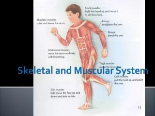
Composed of cells that can contract • Three types of muscle tissue: • Skeletal • Smooth • Cardiac Muscle Tissue
Moves the bones in your trunk, limbs, and face Skeletal Tissue
Handles body functions you can’t control Smooth Tissue
Heart muscle; pumps blood through body Cardiac Tissue
Contains cells that receive and transmit messages in the form of electrical impulses • Neurons: specialized cells that send and receive messages throughout the body • Make up your brain, spinal cord, and nerves • Sense changes in the internal and external environment • Cause the body to move in response Nervous Tissue
Consists of layers of cells that line or cover all internal and external body surfaces • Formed from cells that are tightly bound • Found in various thicknesses and arrangements • Ex: line blood vessels, outer skin Epithelial Tissue
Binds, supports, and protects structures in the body • Most abundant and diverse • Include bone, cartilage, tendons, fat, and blood • Characterized by cells embedded in fluid called matrix • Matrix an be solid, semisolid, or liquid Connective Tissue
Organ: consists of various tissues that work together to carry out a specific function Organ system: groups of organs that interact Organs and Organ Systems
Body cavities: compartments where organs and organ systems are housed • Protect internal organs from injuries and permit organs to move while being supported Body Cavities
Five main body cavities: • Cranial • brain • Spinal • spinal cord • Thoracic cavity • heart, esophagus, respiratory system organs • Abdominal cavity • Digestive system organs • Pelvic cavity • Reproductive and excretory system organs Body cavities
Section 2 Skeletal System
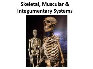
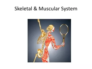
Human skeleton is composed to two parts: • Axial skeleton • Skull, ribs, spine, sternum • Appendicular skeleton • Arms, legs, scapula, clavicle, pelvis The Skeleton
Bones provide a rigid framework against which muscles can pull, give shape and structure to the body, and support and protect internal organs • Bones store minerals • Internal portion of many bones produce red blood cells, platelets, and some white blood cells Bone function and Structure
Bones make up less than 20% of the body’s mass They are moist, living tissues Bone function and Structure
Consists of a porous central cavity surrounded by a ring of dense material • Periosteum: tough membrane that covers bone • Contains a network of blood vessels Long Bone Structure
Most bones develop from cartilage • Ossification: the process by which cartilage is slowly replaced by bone as a result of the deposition of minerals • Some bones develop directly into hard bone without forming cartilage first Bone Development
Bones continue to develop after birth • Bone cells gradually replace the cartilage in long bones of limbs (arms and legs) • Bone elongation takes place near the end of long bones at the epiphyseal plate • Composed of cartilage cells • Growth continues until bone has replaced all of the cartilage in the epiphyseal plate Bone Elongation
Epiphyseal plate Region where cartilage cells divide, enlarge, and push older cells to the middle of the bone Bone Elongation
Joint: the place where two bones meet • Three major types of joints: • Fixed • Semimovable • Movable Joints
Fixed joints prevent movement • Found in the skull where they permit no movement of those bones • Small amount of connective tissue in a fixed joint also helps absorb impact to prevent the bones from breaking Fixed Joint
Semimovable joints permit limited movement • Found in the vertebral column; allow the body to bend and twist • Also found in the rib cage; connect the upper ten pairs of ribs to the sternum Semimovable Joints
Movable joints enable the body to perform a wide range of movements and activities • Include: • Hinge • Ball-and-socket • Pivot • Saddle • Gliding Movable joints
Hinge joint: found in the elbow Ball-and-socket: shoulder joint Pivot joint: formed between the top two vertebrae in the neck Saddle: found at the base of each thumb Gliding: fount between the small bones of your foot Movable Joints
Joints are subjected to a great deal of pressure and stress, but their structure is well suited to meet these demands • Bones that come into contact are covered with cartilage • Protects against friction Joint Structure
Ligaments: tough bands of connective tissue • Hold the bones of the joint in place • Synovial fluid: lubricating substance that helps protect the ends of bones from damage by friction Joint Structure
Knee joint is the most susceptible because it carries the body’s weight Joint Structure
Arthritis: used to describe disorders that cause painful, swollen joints • Two forms of arthritis: • Rheumatoid arthritis: develops when the immune system begins to attack body tissues • Osteoarthritis: degenerative joint disease in which cartilage becomes thinner and rougher, so bones rub against each other Joint Structure
Section 3 Muscular System
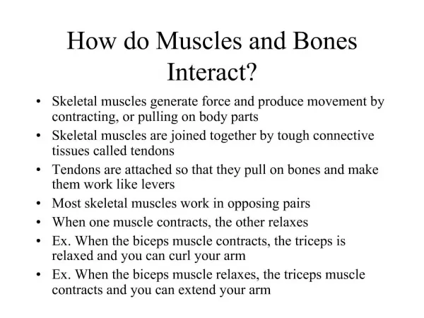
Muscle: an organ that can contract in a coordinated fashion • Includes: • Muscle tissue • Blood vessels • Nerves • Connective tissue Muscle Types
Skeletal muscle is responsible for moving parts of the body • Made of muscle fibers (elongated cells) • Each muscle fiber contains many nuclei and is crossed by light and dark stripes (called striations) • Skeletal muscle fibers are grouped into fascicles (dense bundles) • A group of fascicles are bound together to form a muscle Muscle Types
Voluntary muscles: muscles you can control • Involuntary muscles: muscles you can’t control • Smooth muscle • Makes up the walls of the stomach, intestines, blood vessels, and other internal organs • Cells are spindle-shaped, have a single nucleus, and form sheets • Lacks striations • Fibers surrounded by connective tissue Muscle Types
Cardiac muscle • Make up the walls of the heart • Muscle is striated • Involuntary muscle • Each cell has one nucleus Muscle Types
A skeletal muscle fiber is a single, multinucleated muscle cell • Made up of hundreds or thousands of muscle fibers • Cells are soft and easy to injure • Covered by connective tissue Muscle Structure