Download
1 / 20
200 likes | 284 Views
Explore the parts and characteristics of the large intestine, covering the surface anatomy, peritoneal relationships, arterial and nerve supply. Learn about the anatomy of specific parts like cecum, appendix, colon, rectum, and anal canal.

E N D
ANATOMY OF THE LARGE INTESTINE Dr. Jamila El-Medany Dr. Ahmed Fathalla Ibrahim
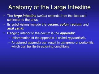
OBJECTIVES At the end of the lecture, students should: • List the different parts of large intestine. • List the characteristic features of colon. • Describe the anatomy of different parts of large intestine regarding: the surface anatomy, peritoneal covering, relations, arterial & nerve supply.
Parts of Large Intestine • CECUM • APPENDIX • ASCENDING COLON • TRANSVERSE COLON • DESCENDING COLON • SIGMOID COLON • RECTUM • ANAL CANAL Abdomen ABDOMEN PELVIS Pelvis PERINEUM Perineum
Characteristics of COLON(NOT FOUND IN RECTUM & ANAL CANAL • Taeniae coli: (3) longitudinal muscle bands 2.Sacculations (Haustra): Because the Taeniae coli are shorter than large intestine 3. Epiploic Appendices :Short peritoneal folds filled with fat
Peritoneal Covering • PARTS WITH MESENTERY: • Transverse colon • Sigmoid colon • Appendix • Cecum • RETROPERITONEAL PARTS: • Ascending colon • Descending colon 3. Upper 2/3 of rectum
Peritoneal Covering PARTS DEVOID OF PERITONEAL COVERING: • Lower 1/3 of rectum • Anal canal Rectum Anal canal
Anterior Relations of (CECUM – ASCENDING & DESCENDING COLONS) • Greater omentum • Coils of small intestine • Anterior abdominal wall
Posterior Relations (CECUM – ASCENDING & DESCENDING COLONS) Cecum: Psoas major Iliacus Ascending colon: Iliacus Quadratuslumborum Right kidney. Descending colon: Left kidney Quadratuslumborum Iliacus Quadratus lumborum
COLIC FLEXURES Position: higher Angle: more acute Hepatic flexure Splenic flexure
Relations of Transverse Colon Anterior: greater omentum, anterior abdominal wall Posterior:2nd part of duodenum , pancreas & superior mesenteric vessels.
Relations of Transverse Colon Superior:liver, gall bladder, stomach Inferior: coils of small intestine
APPENDIX • Surface anatomy: • the base of appendix is marked by Mc’Burney’s point: • A point at the junction of lateral 1/3 & medial 2/3 of a line traced from right anterior superior iliac spine to umbilicus
APPENDIX Opening: At posteromedial aspect of cecum, 1 inch below ileo-cecal junction Positions: 1.Retrocecal :(most common) 2.Pelvic 3.Subcecal 4.Preilieal 5.Postileal:least common (4) (5) (1) (2) (3)
RECTUM Beginning:as a continuation of sigmoid colon at level of S3. Termination: continues as anal canal, one inch below & in front of tip of coccyx.Its end is dilated to form the rectal ampulla. Length: 13 cm(5 inches)
Relations of Rectum in Pelvis MALE PELVIS Anterior:seminal vesicles, posterior surfaces of urinary bladder & prostate gland Posterior: sacrum, sacral plexus & coccyx FEMALE PELVIS Anterior:posterior wall of vagina Posterior:sacrum , sacral plexus & coccyx R R
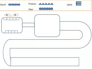
Relation Between Embryological Origin of GUT& its Arterial Supply
VENOUS DRAINAGE OF GUT • The veins of the gut form the tributaries of the portal vein which enters the liver and drains into the portal circulation.
Lymph drainage of Gut • The lymph vessels follow the arteries. • Ultimately, all the lymph is collected at the Preaortic lymph nodes (Superior & Inferior mesenteric).
RELATION BETWEEN EMBRYOLOGICAL ORIGIN & NERVE SUPPLY • Origin:Midgut (endoderm) • Nerve supply:(Autonomic): • Sympathetic + Vagus • Origin:Hindgut (endoderm) • Nerve supply: (Autonomic): • Sympathetic + pelvic splanchnic nerves • Origin: ectoderm (lower 1/3 of anal canal) • Nerve Supply: Somatic(inferior rectal)