Download
1 / 47
480 likes | 822 Views
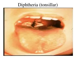
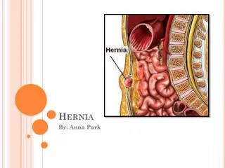
Guirish Solanki Birmingham Children’s Hospital Birmingham, UK. Chiari like Tonsillar Hernia & A novel Management Strategy. Radiological Definitions. 1985 - Aboulezz et al. Tonsils tip up to 3 mm below the Foramen Magnum are normal. In Chiari I the hernia exceeds 5 mm.

E N D
Guirish Solanki Birmingham Children’s Hospital Birmingham, UK Chiari like Tonsillar Hernia&A novel Management Strategy
Radiological Definitions • 1985 - Aboulezz et al. • Tonsils tip up to 3 mm below the Foramen Magnum are normal. • In Chiari I the hernia exceeds 5 mm. • 1986 - Barkovich et al. • The limit is 5 mm below the Foramen Magnum.
Asymptomatic Chiari I 14% of patients asymptomatic Syringomyelia and osseous anomalies in only asymptomatic patients… Tonsillar hernia avg 11.4 mm Incidental in 50% 22% clinical worsening 14% progressed to surgery
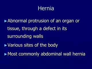
Craniosynostosis & Hindbrain Hernia(a.k.a. Chiari Malformations) Risk Factors for development of Chronic Hindbrain Hernia • Premature fusion of skull vault & skull base sutures • congenital anomalies of the cerebellum & brain stem • Raised intracranial pressure • Venous hypertension • Hydrocephalus • Pfeiffer’s syndrome 50% • Crouzon’s syndrome 70% • Oxycephaly 75% • Kleeblattschädel deformity 100% • Apert’s <5%
MRI Appearance of Tonsillar Hernia in CS • Peglike tonsils • Effacement of CSF space at Foramen Magnum • Flattened Occipital Bone • Steep tentorium – nearly vertical • Venous engorgement • Cervico-medullary kink • “Standing-up” cerebellum
Normal Situation Obtuse angle Tent Pons Sitting Cerebellum Foramen Magnum CSF Flow Spinal cord
Chiari changes Acute angle Tent Small Posterior Fossa Flattened Pons Standing Cerebellum Flat Pons Kinking of cervico-medullary junction Foramen Magnum Herniation of tonsils Chiari with loss of CSF Spinal cord
Surgery in Chiari • Current approach for Chiari I and II • Foramen Magnum Decompression craniectomy • With or without C1-C2 laminectomy • With or without Dural opening • With or without Arachnoid Opening • With or without dural / arachnoid closure • With or without Tonsil resection • With or without Duroplasty • With or without bone replacement
Paediatric Foramen Magnum Dimensions in the Chiari malformations and Syringomyelia: A comparative review R. Vemaraju D. Rodrigues, P.Davies*, N. Furtado, G. Solanki Department of Paediatric Neurosurgery and *R&D Diana, Princess of Wales Children’s Hospital Birmingham Children’s Hospital NHS Foundation Trust 224 MRI Scans
The Foramen Magnum in Chiari Largest increases seen in Sagittal diameter and Surface Area in Chiari I
Rapid Pan-synostosis Progression Progressive coronal, sagittal, metopic & lambdoid suture synostosis. Progressive increase in fingerprint impressions (Copper-beaten appearance) Clinical evidence of raised intracranial pressure Crouzon’s syndrome Age = 1 week Crouzon’s syndrome Age = 2 months Crouzon’s syndrome Age = 5 months
Approaches: Under 1 year of age Rapid Progression with Chiari & ventriculomegaly Kleeblattschädel deformity MRI age 2 months • Severe pansynostosis • Brachyturricephaly • Small posterior fossa • Ventriculomegaly • Hindbrain hernia • No fixation • Supra-tentorial Augmentation alone • Posterior Augmentation alone • Supra-tentorial & Occipital Augmentation
If the problem is at the back of the head, operate at the back of the head Supra Regional Craniofacial Unit Birmingham, UK Posterior Release 1984-2003 Posterior Augmentation 2003-2006 Posterior Distraction 2006+ → → www.bch.nhs.uk/departments/craniofacial
Radiological Parameters CERVICOMEDULLARY KINK “STANDING-UP “ CEREBELLUM CSF FLOW AT CCJ TONSILLAR DESCENT SYRINX • CSF DISTRIBUTION IN SULCI • VENOUS HYPERTENSION • TENTORIAL ANGLE • BOWING OF CORPUS CALLOSUM • VENTRICULOMEGALY
5 year old with bi-lambdoid synostosis, ventriculomegaly and hindbrain hernia. Symptomatic with raised ICP. There is less tonsilar descent when compared to pre Fixed Calvarial Augmentation. CSF is clearly seen surrounding the cord and tonsils at the cranio-vertebral junction. No syrinx in the upper cervical cord is now noted.
Results • Reduced density of the occipital cortex / lobe • Despite posterior distraction frontal expansion occurred • Improved CSF distribution • Improved Callosal and ventricular shape. • Reduction of raised ICP • Chiari malformation • CSF Flow? Pre-Op Post-Op
Pre-op 1 year post-op 2 years post-op Reduced Chiari Improved csf flow
POST-OP 1 YEAR PRE-OP
PRE-OP POST-OP 4 YEARS
PRE-OP POST-OP 4 YEARS
PRE-OP POST-OP 4 YEARS
Surgical results Overall 88%(15/17) patients showed improvement in the radiological criteria
Patients withTonsillar Hernia 92%(12/13) Patients with Tonsillar Hernia showed improvement in radiological criteria
Follow-up & Improvement • Overall Radiological criteria • Improvement noted (12) 27 months • No improvement (1) 7.4 months • Tonsillar Hernia • Reduced in 9/13 cases 69% • Lambdoid Synostosis 25 months • Others 31 months
Complications • Wound complications • Bony non-union • Buckling of resorbable plates • Loosening of screws and unstable construct Reasons for Developing Posterior Distraction
Posterior Calvarial Distraction in Multiple Suture Synostosis Latency Distraction Consolidation
Tonsillar Hernia in Craniosynostosis Restricted Skull Expansion during rapid brain growth Volume reduction / distribution Some compaction of brain Outer cortex (lays bone) at sutures = Ridging Inner cortex (looses bone) remodells = Copper beaten appearance Reduction in Ventricular size or Increased ventricles / CSF space (if obstruction to CSF e.g AS) Transient Increase in ICP • Tonsillar herniation • Ocipital lobe is pushed behind tent • Posterior fossa crowding • Cerebellum pushed forwards • Brainstem kinks forwards Loss of CSF at CCJ Syrinx
Conclusion • Fixed Posterior Calvarial Augmentation • Is effective in management of raised ICP in CS associated with craniocephalic mismatch • Improves radiological appearance in a number of features that suggest the brain has more space around it. • Reduction in Syrinx size