Download
1 / 25
310 likes | 1.07k Views
Right Laparoscopic Adrenalectomy. University of Kentucky Minimally Invasive Surgery Elective. Indications for Laparoscopic Approach . Adrenocortical tumors related to: Cushing’s Disease Conn’s Disease Virilization of females Feminization of males. Pheochromocytomas

E N D
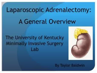
Right Laparoscopic Adrenalectomy University of Kentucky Minimally Invasive Surgery Elective
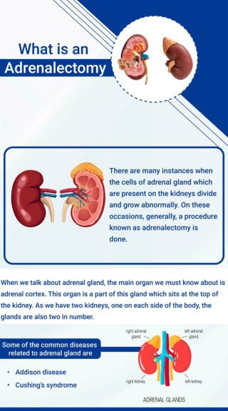
Indications for Laparoscopic Approach Adrenocortical tumors related to: • Cushing’s Disease • Conn’s Disease • Virilization of females • Feminization of males Pheochromocytomas Incidentalomas (of sizes greater than 3-4 cm)
Contraindications for Laparoscopic Approach • Adrenal tumors greater than 8-10 cm • Adrenal Carcinoma • Intracranial hypertension and coagulation issues • These are contraindications in all laparscopicsugery. • Surgical history of kidney of liver • This is due to an increased risk of adhesions, which would not allow for a transperitoneal approach to be utilized.
Procedure Positioning (Patient) • The patient is placed in a left lateral decubitus position, with the table flexed at the midline. This opens up the operating field. • A cushion is often placed under the left flank of the patient. • The legs of the patient are flexed in order to avoid neuropathy of the lower extremities.
Procedure Positioning (Surgical Team) • Both the primary surgeon and the assisting surgeon stand on the abdominal side of the patient. • The assisting nurse stands opposite of the surgeons. • The anesthesiologist stands at the head of the table.
Procedure Positioning (Equipment) • The anesthetic equipment is placed at the head of the bed. • The instrument table is placed at the foot of the bed next to the nurse. • There are monitors on both sides of the operating table.
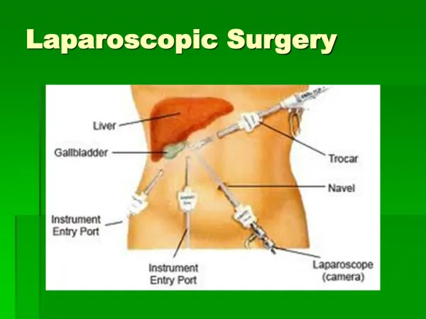
Port Placement • There are four 10 mm trocars utilized in the right adrenalectomy. • There is one placed at the anterior axillary line, under the costal margin. • Another trocar is placed at the mid-clavicular line. • There two remaining trocars are placed one either side of the previously placed trocars, still parallel with the costal margin.
Instruments Required • 30 Degree Laparoscope • DeBakey Grasper • Harmonic Ace curved shears • Laparoscopic scissors • Hook Cautery (sometimes used instead of Harmonic) • Clip Applier • Suction-Irrigation Device • Extraction Bag
Procedure • Mobilization of the liver: • The liver is retracted with the use of a snake retractor. When doing this, compression of the gallbladder should be avoided. • Once this has been accomplished, the subhepatic peritoneum is dissected. This will free the triangular ligament of the liver.
Procedure • The dissection of the subhepatic peritoneum should allow for the surgeon to see the vena cava and the un-dissected adrenal gland behind it. • Identification of the main adrenal vein: • The medial aspect of the gland should dissected towards the vena cava. • The right main adrenal vein should be seen emptying into the vena cava. • Typically, 3 clips are applied, 2 distally and 1 proximally.
Procedure • In approximately 10% of cases, there is an accessory adrenal vein that also requires ligation. • If present, it can be seen connecting to the right suprahepatic vein. • It should also be clipped and ligated.
Procedure • Identification and ligation of the adrenal arteries: • First, the middle adrenal artery should be ligated. It should be seen originating from the aorta. • Next, the superior adrenal artery should be ligated. The adrenal gland should be retracted caudally, making it easier to observe this artery stemming from inferior phrenic artery. • Last, the inferior adrenal artery should be ligated. In reflecting the adrenal gland rostrally, this artery can typically be seen branching off of the renal artery.
Procedure • Once all arteries and veins have been clipped and ligated, complete dissection of the superior, medial, and inferior portions of the gland can take place. • Following this, an extraction bag is utilized to carefully remove the gland from the patient.
Potential Complications • Damage to Liver • Such injury can occur during retraction or during dissection itself. • Damage to Vena Cava • This is the leading cause of conversion to open surgery. • If the lesion is less than 2 mm in size, then it is quite possible that compression and coagulating agents will suffice.
Post-operative Care • The patient may ambulate on the day of surgery. • By the night of the surgery, the patient is allowed fluids. • On the first post-operative day, the patient is allowed to consume solid food. • Release from hospital typically occurs on the 2nd or 3rd post-operative day.
Difficulty of the Procedure • Because of the retroperitoneal location of the adrenal glands, dissection of peritoneum and other fascia often account for the majority of operation time. • This extensive dissection can be a hassle. To compound the problem, a survey discovered that, on average, general surgery residents only received exposure to 1.5 adrenalectomies during their residency.
Differences of Right and Left Adrenalectomies • Right adrenalectomies tend to considered more difficult than left adrenalectomies. • Common thoughts that support this: • Retrocaval location of right adrenal gland • Difficulty of handling the short main adrenal vein that drains into the vena cava.
Study Concerning Differences in Right and Left Adrenalectomies • To investigate this matter, a retrospective study of 163 laparoscopic adrenalectomies was performed. • The study was performed over an 8-year period, following 27 surgeons at 9 Southern California Kaiser Permanente Hospitals. • 109 of the surgeries were left adrenalectomies, while 54 were right adrenalectomies.
Outcomes • Blood Loss • The average estimated blood loss of the left adrenalectomies was 113 mL, ranging from 2 to 3000 mL. • The average estimated blood loss of the right adrenalectomies was 84 mL, ranging from 10 to 700 mL. • This was shown to not be statistically different. • Procedural Time • This however, was statistically different. • Procedural time from left adrenalectomies was, on average, 31 minutes longer than right adrenalectomies.
Plausible Explainations: • Proximity to tail of pancreas • There was an 8% rate of distal pancreatic injury reported. • Complexity of splenic vasculature • Required dissection of left renal hilum • Less mobilization is required for right colon than for the splenic flexure.
Retroperitoneal Approach? • In a study comparing retroperitoneal approach with the transperitoneal approach, it was found that the operation time for the retroperitoneal approach ranged from 290 to 330 minutes. • In comparison, the transperitoneal approach averaged 140 minutes in duration. • Why? • It was found that maneuvering of the surgical tools proved difficult because of a smaller operating field. • In addition, less of the adrenal gland is exposed in this approach.
References • Laparoscopic Right and Left Adrenalectomies: Surgical Endoscopy. (http://www.ncbi.nlm.nih.gov/pubmed/8703150) • Differences in Right and Left Adrenalectomies: Journal of the Society of Laparoendoscopic Surgeons. (http://www.ncbi.nlm.nih.gov/pmc/articles/PMC3041033/) • Laparoscopic Right Adrenalectomy:WebSurg. (http://chapters.websurg.com/technique/index.php?doi=ot02en211&s=12&k=2) • Images from Adrenal Surgery. (http://www.endocrinesurgery.net.au/laparoscopic-adrenalectomy/)