Download
1 / 24
270 likes | 711 Views
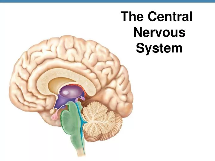
The Central Nervous System. Cerebral hemisphere. Outline of diencephalon. Midbrain. Cerebellum. Pons. Medulla oblongata. Spinal cord. (a) Week 13. Figure 7.12. Features of the Cerebrum. Anterior. Surface markings Gyri = ridges Sulci = shallow grooves Fissures = deep grooves Lobes

E N D
Cerebral hemisphere Outline of diencephalon Midbrain Cerebellum Pons Medulla oblongata Spinal cord (a) Week 13 Figure 7.12
Features of the Cerebrum Anterior • Surface markings • Gyri = ridges • Sulci = shallow grooves • Fissures = deep grooves • Lobes • Frontal • Parietal • Temporal • Occipital Longitudinal fissure Left cerebral hemisphere Right cerebral hemisphere Posterior
Precentral gyrus Central sulcus Postcentral gyrus Frontal lobe Parietal lobe Occipital lobe Temporal lobe Cerebellum Fissure Gyrus Cortex (gray matter) Sulcus White matter (a) Figure 7.13
The Principal Parts of the Adult Brain Cerebrum Diencephalon Cerebellum Brain stem (b) Adult Brain Figure 7.12
Cerebral Cortex • Thin superficial layer of gray matter • Deeper white matter composed of fiber tracts carrying impulses to or from cortex • Corpus callosum- connects cerebral hemispheres
Diencephalon • Enclosed by cerebral hemispheres • Major structures of diencephalon: • Thalamus • Hypothalamus • Epithalamus 11
Thalamus • Relay station for sensory impulses passing up to sensory cortex • Encloses the 3rd ventricle
Hypothalamus • Below thalamus • Floor of diencephalon • Pituitary hangs from hypothalamus • Important autonomic nervous center for: • Body temperature • Metabolism • Water balance
Epithalamus • Forms roof of 3rd ventricle • Pineal body- part of endocrine system • Choroid plexus – capillary beds which form CSF
Thalamus Epithalamus Hypothalamus Pituitary Gland
The Brain Stem • Midbrain • Pons • Medulla Oblongata
The Midbrain • Superior portion of brainstem • Cerebral aqueduct passes through to connect 3rd ventricle above and 4th ventricle below • Anteriorly composed of tracts called cerebral peduncles that • Convey ascending & descending impulses • Dorsally placed are corpora quadrigemina that are • Reflex centers for hearing, vision
Cerebral Aqueduct Coropora Quadrigemina Cerebral Peduncles
11 The Pons • Rounded structure just below midbrain • Mostly fiber tracts that bridge areas of brain • Contains nuclei involved in the control of breathing Pons
Medulla Oblongata • Inferior portion of brain stem • Many important fiber tracts • Contains nuclei that regulate vital visceral activities • Heart rate • Blood pressure • Breathing • Swallowing, etc. Medulla Oblongata
11 Cerebellum • Functions: • Precise timing for skeletal muscle activity • Controls balance & equilibrium
Meninges • Functions: • Cover and protect the CNS • Contain cerebrospinal fluid (CSF) • Form partitions in the skull • Three layers • Dura mater • Arachnoid mater • Pia mater
Skin of scalp Periosteum Bone of skull Outer Dura mater Inner Superior sagittal sinus Arachnoid mater Pia mater Subdural space Subarachnoid space Figure 7.16
Meninges • Dura Mater (Strongest, Outer layer) • Two layers (fibrous CT); separate to form dural sinuses • Dural septa – inward folds of dura mater • Falx cerebri—in longitudinal fissure • Falx cerebelli— separates cerebellum in half • Tentorium cerebelli—horizontal; separates cerebellum and cerebrum
Falx cerebri Crista galli Of ethmoid bone Tentorium cerebelli Falx cerebelli (a) Dural septa
Meninges • Arachnoid Mater- Middle layer • Weblike • Contains CSF & blood vessels • Arachnoid villi extend into dural sinus; allow CSF reabsorption • Pia Mater -Innermost layer • Delicate vascularized CT that clings tightly to the brain
Ventricles of the Brain • Connected to one another and to central canal of spinal cord • Two C-shaped lateral ventricles in the cerebral hemispheres • Third ventricle in diencephalon • Fourth ventricle is dorsal to pons
Lateral ventricle Interventricular foramen Lateral aperture Third ventricle Cerebral aqueduct Fourth ventricle Central canal (a) Anterior view (b) Left lateral view Figure 7.17