Download

1 / 78
850 likes | 1.1k Views
Gestational Trophoblastic Neoplasia (GTN). Zohreh Yousefi Professor of Obstetrics and Gynecology, Fellowship of Gynecology Oncology , Ghaem Hospital, website: www.zohrehyousefi.com. Danforth's Williams Obstetrics, 23e

E N D
Gestational Trophoblastic Neoplasia (GTN) ZohrehYousefi Professor of Obstetrics and Gynecology, Fellowship of Gynecology Oncology , Ghaem Hospital, website: www.zohrehyousefi.com
Danforth's Williams Obstetrics, 23e B erek and Hacker's Gynecologic Oncology Up To Dat GESTATIONAL TROPHOBLASTIC DISEASE PATHOGENESIS DIAGNOSIS MANAGEMENT GESTATIONAL TROPHOBLASTIC NEOPLASIA TREATMENT SUBSEQUENT PREGNANCY
Gestational trophoblastic disease (GTD) is term group of tumors with abnormal trophoblast proliferation produce human chorionic gonadotropin (hCG)
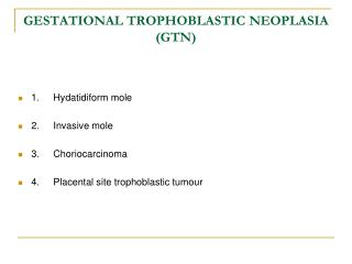
GTD histologically is divided into benign hydatidiform moles ( complete and partial) Malignant Invasive mole
Non -molar trophoblastic neoplasms • Choriocarcinoma • Placental site trophoblastic tumor • Epithelioid trophoblastic tumor
Gestational trophoblastic neoplasia (GTN ) Malignant forms of gestational trophoblastic disease GT N is all GTD except hydatidiform mole Weeks or years following any type of pregnancy But frequently occur after a hydatidiform mole
Hydatidiform mole Microscopic (classic findings) Absence embryonic elements Trophoblastic proliferation (cytotrophoblast and syncytiotrophoblast) Stromal edema and hydropic degeneration Absence of blood vessels
Macroscopic of Hydatidiform mole Hydropic villi Grapelike vesicles filled clear material usually 1 to 3cm diameter proliferation of the trophoblast
Hydatidiform mole Complete mole Partial mole Partial mole Partial mol ( fetal tissue) Grossly placenta a mixture of normal and hydropic villi Fetus Severe growth restriction Multiple congenital anomalies
Risk Factors hydatidiform mole Strongest risk factors are Age and a history of prior hydatidiform mole Both extremes of reproductive age adolescents twofold risk Older than 40 tenfold risk
history of Prior mole • the risk of another mole • Complete mole is 1.5 percent • Partial mole is 2.7 percent • Two prior molar pregnancies • the risk is 23 percent
An ethnic predisposition • Diet (Deficiencies of protein or) • (Vitamin A deficiency) • animal fat • Smoking • Increased paternal age
Pathogenesis Abnormal fertilization process Normal ovum with a duplicated haploid sperm Inactive ovum chromosomes Karyotype 46, XX diploid and result from androgenesis Partial moles triploid karyotype 69, XXX, 69,XXY
Clinical Findings Because universal sonography in prenatal care Typically diagnosed at a mean of 10 weeks • Vaginal bleeding • spotting to profuse hemorrhage • Moderate iron-deficiency anemia
Exaggerated early pregnancy symptoms • Nausea and vomiting ( hyperemesis) • Abdominal cramp
Abnormally enlarged and soft uterus uterine growth Theca-lutein cysts (hCG) 25 to 60% (Torsion, infarction, rupture and hemorrhage) Releases antiangiogenic factors that activate endothelial damage Severe preeclampsia hypoxic trophoblastic mass
All hydatidiform moles secrete hCG Thyrotrophic -like effects of hCG hCG acts a thyrotrophic substance Elevated serum free thyroxine (T4) (TSH) levels to be decreased thyroid hyper –function “thyroid storm”
Diagnosis Amenorrhea followed by irregular bleeding Spontaneous passage of molar tissue High values Serum β-HCG measurement confirming the diagnosis IHC stain positively for p57
Sonography Echogenic uterine mass with anechoic cystic spaces without a fetus or amnionic sac The appearance as “snowstorm
Transvaginal sonogram demonstrating the “ snow storm” appearance.
Mis-diagnosis • Incomplete abortion • missed abortion • Cystic degeneration • uterine leiomyoma
which of the following symptoms will a highly intelligent physician assistant immediately consider hydatidiform mole? • pelvic pain at night during the first trimester • significantly elevated BP in the first trimester • significant bloody vaginal discharge in the first trimester • nausea and vomiting in the first trimester
Management Termination of Molar Pregnancy • Evacuation and Curettage • Hysterectomy (rarely and select cases • no desired future pregnancy ) • Chest radiograph • Initiate effective contraception • OCP or MPA } poor compliance}
Serum hCG levels: 48 hours of evacuation (baseline) Weekly until undetectable Weekly until normal for 3 consecutive weeks monthly until normal for at least 6 consecutive months Median time for resolution is 9 weeks for complete 7 weeks for partial Hysterectomy reduces the incidence of malignant sequelae does not eliminate follow-up
hCG change HM: 84-100 days Spontaneous abortion: 19 days Normal delivery: 12 days Ectopic pregnancy 8-9 days
After molar evacuation risk factors for malignant squeal 15 - 20 % complete moles 1 - 5 % partial moles 1 5% of HM become invasion moles 2.5% progress intochoriocarcinoma
Twin Pregnancy (Normal Fetus and Coexistent Complete Mole) Diagnosis is difficult (early pregnancy ultrasound) A single partial molar pregnancy with abnormal fetus Distinguished
A few cases the diagnosis is not suspected until examination of the placenta following delivery
Amniocentesis ( fetal karyotype ) diploid or triploid If fetal karyotype is normal Major fetal malformations are excluded by ultrasound Chest X-ray performed Serum hCG values If there is no evidence of metastatic disease to allow the pregnancy
Possible risk for developing • Subsequent GTN • Preterm delivery • Preeclampsia • Sever hemorrhage
Persistent GTD : Persistently elevated serum hCG level Irregular vaginal bleeding Persistent theca lute in cysts (2 to 4 months regress spontaneously) Uterine sub involution Risk factors for GTN
Risk factors of GTN Older age β-hCG levels > 100,000 mIU/mL Large uterine size for-gestational age Theca-lutein cysts > 6 cm Earlier recognition and evacuation of molar pregnancies not lower risk neoplasia
Criteria for Diagnosis of Gestational Trophoblastic Neoplasia Criteria for the diagnosis of postmolar GTN 1. Plateau or rise of serum β-hCG level 2. Detectable serum β-hCG level for 6 months or more 3. Histological criteria for choriocarcinoma 4-Irregular bleeding ,uterine sub involution
Plateau of serum β-hCG level (± 10 percent) for four easurements during a period of 3 weeks or longer days 1, 7, 14, 21 Rise of serum β-hCG level > 10 percent during three weekly consecutive , during a period of 2 weeks or more—days 1, 7, 14
Diagnosis Sonography Abdomino pelvic or trans vaginal sonography Radiograph of chest Chest CT scan Brain CT scan or MRI
SPESIAL 1-Selective angiography of abdominal and pelvic or hepatic (if indicated ) 2-Whole body PET Less commonly (occult disease ) 3-Stool guaiac tests If positive test is or if gastrointestinal symptoms be routinely performed in persistent GTN 4- complete radiographic evaluation of the gastrointestinal tract
GTN CLASSIFICATION Invasive Mole Almost all invasive moles arise from partial or complete moles Deep penetration into the myometrium or peritoneum Involvement of vaginal vault
Choriocarcinoma Most common follow a term pregnancy or miscarriage Rapidly growing both myometrium and blood vessels Blood-borne metastases
differentiation between invasive mole and choriocarcinoma if we see villi, it must be invasion mole if we can’t see villi, it is choriocarcinoma
Common Sites for Metastatic Gestational Trophoblastic Tumors
Symptoms • Metastatic symptoms • Profuse vaginal bleeding • Vaginal or cervical metastasis • (bluish nodule in vaginal) • Abdominal pain (intra-abdominal hemorrhage) • Cough, hemoptysis • Headache, nausea, vomit, paralysis or coma • Urologic hemorrhage
Lung metastasis Four principal pulmunary radiologic patterns: • Snowstorm pattern (Alveolar pattern ) • Discrete rounded densities • Plural effusion • Embolic pattern
Brain metastasis • Plasma CSF /hCG level ratio is normally • >60: 1 • In patients with CNS metastases <60: 1 • Falsely lowered plasma CSF /hCG level • First -trimester abortions In the absence of lung or vaginal metastasis Risk of cerebral and hepatic spread is exceedingly low
Generally in GTN Serum hCG levels combined Clinical findings Rather than a histological specimen Diagnose and treat this malignancy
Follow-up of GTN patients β-subunit until hCG Weekly until normal for 3 consecutive weeks monthly until normal for at least 3 consecutive months at 1-month interval for 1 year: at 1- month interval for 2 years in high stage at yearly interval for many years (increased risk of late recurrence)