Download
1 / 48
530 likes | 973 Views
Introduction and classification of glaucoma and Congenital Glaucoma. Dr.Ajai Agrawal , Additional Professor, Department of Ophthalmology, AIIMS, Rishikesh. Acknowledgement. Becker- Schaffer’s Diagnosis and therapy of The Glaucomas (8 th Edition ).

E N D
Introduction and classification of glaucoma and Congenital Glaucoma Dr.AjaiAgrawal, Additional Professor, Department of Ophthalmology, AIIMS, Rishikesh.
Acknowledgement • Becker- Schaffer’s Diagnosis and therapy of The Glaucomas (8th Edition). • Kanski’s Clinical Ophthalmology (8th Edition). • Comprehensive Ophthalmology (A.K.Khurana) (7th Edition).
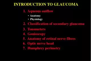
Learning Objectives • At the end of this class the students shall be able to : • Define and classify glaucoma. • Define congenital glaucoma. • Understand the aetio-pathogenesis and clinical features of congenital glaucoma. • Understand the fundamentals of managing congenital glaucoma.
Excessive blinking +/- watering • Hazy cornea • Large corneas
Question • A child presents with watering , photophobia and an enlarged cornea with a diameter of 13mm. Examination of the eye reveals double contoured opacities concentric to the limbus. Which of the following is the most likely diagnosis: • Superficial keratitis • Deep keratitis • Thyroid eye disease • Congenital glaucoma
What is glaucoma ? • The term glaucoma is derived from the Greek word “glaukos” meaning “gray blue” • Second leading cause of blindness worldwide • Third most common cause of blindness in India • Not reversible
Definition of glaucoma • Group of disorders characterized by progressive optic neuropathy resulting in characteristic morphological changes at the optic disc leading to a specific pattern of irreversible visual field defects (with or without a raised IOP).
Primary glaucoma • Open angle glaucoma • Primary Open angle glaucoma • Normal Tension glaucoma • Juvenile Open angle glaucoma • SecondaryOpen angle glaucoma • Steroid induced glaucoma • Pigmentary glaucoma
Primary glaucoma • Angle closure glaucoma • Primaryangle closure glaucoma • Secondary angle closure glaucoma • Swollen lens • Posterior segment tumours • Neovascular glaucoma • Plateau iris syndrome
Childhood glaucoma • Primary congenital glaucoma • Glaucoma associated with ocular abnormalities • Glaucoma associated with systemic abnormalities
Childhood glaucoma-Introduction • Diverse group of disorders • Primary congenital glaucoma- Developmental abnormality of angle of anterior chamber leading to high intraocular pressure(IOP). • Secondarycongenital glaucoma With associated ocular and systemic anomalies
ANGLE OF ANTERIOR CHAMBER - The peripheral recess of anterior chamber is known as the angle of anterior chamber. - It is clinically visualized by gonioscopy. - Starting at the root of iris & progressing anteriorly towards the cornea, the following structures can be identified in a normal angle in an adult : 1) Ciliary body band (CBB) & root of iris 2) Scleral spur (SS) 3) Trabecular meshwork (TM) 4) Schwalbe’s line (SL)
--------- Grade IV III II I 0
Childhood glaucomas • True congenital glaucomas- At birth or during intrauterine period. • Infantileglaucoma- Upto three years of age. • Juvenileglaucoma- After three years of age and upto 35 years of age.
Prevalence and genetic pattern • Sporadic occurrence in most cases (90%) • Autosomal recessive in 10% of cases • Loci linked with congenital glaucoma are 2p21(GLC3A), 1p36(GLC3B) and 14q24(GLC3C) • 60% diagnosed by the age of 6 months and 80% diagnosed within the first year of life
Prevalence and genetic pattern • Bilateral (about 70%) but asymmetrical • Boys are affected slightly more frequently than girls (65%) • Prevalence is 1 in 10,000 births • Chance of second sibling having disease is 3% • Chance of third sibling (of two affected siblings) having disease is 25%
Pathogenesis • Faulty development of angle of anterior chamber from neural crest derived cells (trabeculodysgenesis) • Absence of angle recess with flat/concave iris insertion. • Impaired aqueous outflow • Elevated IOP The normal chamber angle: on the left is a histological cross-section; on the right is a drawing of the same An underdeveloped chamber angle
Clinical presentation Classic triad of • Epiphora • Blepharospasm • Photophobia • Babies rub their eyes • Enlarged eyes • Vision impaired
Corneal signs • Corneal oedema • Corneal enlargement (Corneal diameter>13mm) • Haab’s striae:Descemet’s membrane is not very elasticand stretching may result in small linear/circumferential tearsthat cause a certain degree of corneal opacification.
Clinical presentation • Buphthalmos:Enlargement of the globe as a result of elevated IOP. All segments of the outer eyeespecially the cornea and scleraexpandprincipally at the corneoscleral junction • The anatomic landmarks are displaced. • The anterior chamber is deep Advanced developmental glaucoma with extensive enlargement and scarring of the cornea.
Clinical presentation • Sclera becomes thin and appears blue (due to underlying uveal tissue • Iris- atrophic in later stages • Optic disc- variable cupping • Intraocular pressure(IOP)- raised • Axial myopia- due to increased axial length of eyeball
Examination under anaesthesia • Mandatory in all cases • Includes : • Measurement of IOP – Perkins tonometer/Tonopen (Normal 10-21 mm Hg) • Measurement of corneal diameter – by callipers (Normal 9.5mm-10.5mm)
Examination under anaesthesia • Slit lamp examination- with portable slit lamp • Ophthalmoscopy- to evaluate optic disc Asymmetric disc cupping in a child with developmental glaucoma. (A) Note steep-walled cup. This is typical of glaucomatous cupping in the elastic infant eye. (B) The left eye has no cupping.
Examination under anaesthesia • Direct Gonioscopy– to examine angle of anterior chamber • Koeppe’sgonioscopy lens is preferable • Angle is open but immature in congenital glaucoma
Differential Diagnosis • Hazy/Cloudy cornea---- • STUMPED (Sclerocornea, Trauma, Ulcer, Metabolic disorders, Peter’s anomaly, Endothelial dystrophy) • Watering and intolerance to light----- Congenital Naso Lacrimal Duct obstruction keratitis, conjunctivitis • Optic cupping ---- disc coloboma, hypoplasia, physiological cupping • Corneal enlargement-- megalocornea, high myopia • Descemet’sbreaks --- Forceps delivery ,birth trauma
Management • Glaucoma surgery is the primary option • Medications are not very effective • Role of medical management is temporary, till surgery is taken up. • Beta blockers (Timolol), hyperosmotic agents(Mannitol), carbonic anhydrase inhibitors (acetazolamide/dorzolamide) • Miotics and Alpha-2 agonists are not used in children.
Approach to management Goniotomy/Trabeculectomy/Combined Trabe-Trab Surgical outcome? EUA after 3-4 weeks IOP not controlled IOP controlled Add medical therapy Evaluation after 3 months If IOP not controlled repeat Trab ± MMC Normal IOP Evaluation after 3 months Uncontrolled Controlled Poor prognosis FU every 3 months VISUAL REHABILITATION Consider Drainage implant Cyclodestruction Record IOP, CDR, VA Axial length, VF (if possible)
Goniotomy • Safe procedure when performed skilfully. • Performed with direct visualization of trabecular meshwork • Aims to transect Schlemm’s canal by ab-interno approach • Incises only superficial trabecular tissues, necessary to cure this disease
Trabeculotomy • Ab-externo trabeculotomy has good success rates.
Trabeculotomy with trabeculectomy • Most commonly performed surgery in India • Easy adaptability • Safe and successful • Suitable in compromised corneas • More predictable results
Steps of Trabeculectomy with trabeculotomy Scleral flap Ds sDissectionupto grey limbus Trabecular meshwork cut Diffuse subconjunctival bleb
Role of antimetabolites in paediatric glaucoma • Significantly more complications associated with the use of Mitomycin(MMC) in paediatric glaucomas • Thin, avascular filtering blebs • Wound leakage • Choroidal detachment • Bleb related endophthalmitis
Options for refractory glaucoma ? • Glaucoma Drainage Devices • Cyclo-destruction
What is a Glaucoma Drainage Device? Glaucoma drainage devices (GDDs) create an alternate aqueous pathway from the anterior chamber (AC) by channeling aqueous out of the eye through a tube to a subconjunctival bleb. This tube is usually connected to an equatorial plate under the conjunctiva.
Cyclodestructive procedures • Cyclocryotherapy • Cyclophotocoagulation • Transscleral • Transpupillary • Endoscopic
CYCLO CRYOTHERAPY TREATMENT OF 1950 BIETTI
Lasers relatively safer energy • Trans-scleral route • Direct application to ciliary • epithelium • Trans pupillary
Trans-scleral route diode laser 810 nm wave length Penetrates through sclera Contact delivery through fibre optic cable Diode laser is preferred Melanin in the ciliary epithelium better absorbs this wavelength Causes more targeted destruction with less inflammation
VISUAL REHABILITATION • Correction of refractive error • Management of media opacities • Amblyopia therapy to achieve binocular stereoscopic vision
VISUAL REHABILITATION • Low vision aids • Telescopes (hand-held or spectacle-mounted) • Hand or pocket magnifiers (2× to 3×)
CONCLUSIONS • Glaucoma is a group of disorders characterized by progressive optic neuropathy. • Early diagnosis and prompt treatment can preserve vision. • All children with suspected childhood glaucoma should be examined under anaesthesia. • Mainstay of management of childhood glaucoma is surgery • Visual rehabilitation and counseling of the parents of the child is as important as IOP control.
Question • Identify the abnormality marked by arrow. • Which structure is involved? • What type of slit lamp illumination is used in the photograph? • Mention one differential diagnosis of the condition


![[Glaucoma] Classification](https://cdn1.slideserve.com/2649686/glaucoma-classification-dt.jpg)