Download
1 / 1
10 likes | 178 Views
Rodent Islet Amyloid Polypeptide Inhibits Amyloid Formation by Human Islet Amyloid Polypeptide: Implications For the Design of Inhibitors and For Animal Models of Diabetic Amyloid Ping Cao 1 , Fanling Meng 1 and Daniel P. Raleigh 1,2,3

E N D
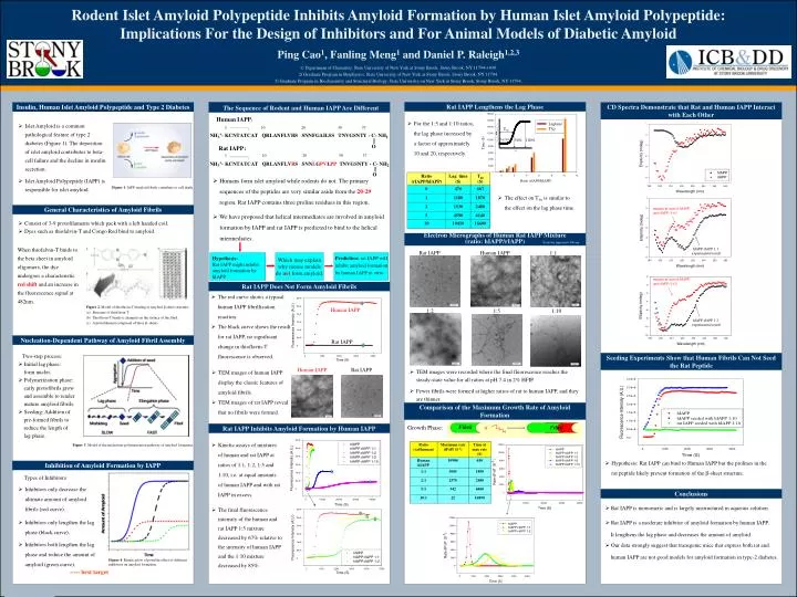
Rodent Islet Amyloid Polypeptide Inhibits Amyloid Formation by Human Islet Amyloid Polypeptide: Implications For the Design of Inhibitors and For Animal Models of Diabetic Amyloid Ping Cao1, Fanling Meng1 and Daniel P. Raleigh1,2,3 1) Department of Chemistry, State University of New York at Stony Brook, Stony Brook, NY 11794-3400. 2) Graduate Program in Biophysics, State University of New York at Stony Brook, Stony Brook, NY 11794. 3) Graduate Program in Biochemistry and Structural Biology, State University on New York at Stony Brook, Stony Brook, NY 11794. Rat IAPP Lengthens the Lag Phase Insulin, Human Islet Amyloid Polypeptide and Type 2 Diabetes CD Spectra Demonstrate that Rat and Human IAPP Interact with Each Other The Sequence of Rodent and Human IAPP Are Different • Islet Amyloid is a common pathological feature of type 2 diabetes (Figure 1). The deposition of islet amyloid contributes to beta-cell failure and the decline in insulin secretion. • Islet Amyloid Polypeptide (IAPP) is responsible for islet amyloid. Human IAPP: • For the 1:5 and 1:10 ratios, the lag phase increased by a factor of approximately 10 and 20, respectively. 1 10 20 30 37 T50 Lag time 50% 100% Rat IAPP: 1 10 20 30 37 • Humans form islet amyloid while rodents do not. The primary sequences of the peptides are very similar aside from the 20-29 region. Rat IAPP contains three proline residues in this region. • We have proposed that helical intermediates are involved in amyloid formation by IAPP and rat IAPP is predicted to bind to the helical intermediates. Figure 1. IAPP amyloid fibrils contribute to cell death. • The effect on T50 is similar to the effect on the lag phase time. General Characteristics of Amyloid Fibrils numerical sum of hIAPP and rIAPP (1+1) • Consist of 3-9 protofilaments which pack with a left handed coil. • Dyes such as thiofalvin-T and Congo Red bind to amyloid. Electron Micrographs of Human Rat IAPP Mixture (ratio: hIAPP/rIAPP) Scale bar represents 100 nm When thiofalvin-T binds to the beta sheet in amyloid oligomers, the dye undergoes a characteristic red shift and an increase in the fluorescence signal at 482nm. hIAPP:rIAPP 1:1 experimental result Rat IAPP Human IAPP 1:1 Which may explain why mouse models do not form amyloid. Hypothesis: Rat IAPP might inhibit amyloid formation by hIAPP. Prediction: rat IAPP will inhibit amyloid formation by human IAPP in vitro. numerical sum of hIAPP and rIAPP (1+2) Rat IAPP Does Not Form Amyloid Fibrils • The red curve shows a typical human IAPP fibrillization reaction. • The black curve shows the result for rat IAPP, no significant change in thioflavin-T fluorescence is observed. • Figure 2. Model of thioflavin-T binding to amyloid β-sheet structure. • Structure of thioflavin-T. • Thioflavin-T binds to channels on the surface of the fibril. • A protofilament composed of three β–sheets. Human IAPP 1:2 1:5 1:10 hIAPP:rIAPP 1:2 experimental result Nucleation-Dependent Pathway of Amyloid Fibril Assembly Rat IAPP Seeding Experiments Show that Human Fibrils Can Not Seed the Rat Peptide • Two-step process: • Initial lag phase: form nuclei. • Polymerization phase: early protofibrils grow and assemble to render mature amyloid fibrils. • Seeding: Addition of pre-formed fibrils to reduce the length of lag phase. Human IAPP Rat IAPP • TEM images of human IAPP display the classic features of amyloid fibrils. • TEM images of rat IAPP reveal that no fibrils were formed. • TEM images were recorded where the final fluorescence reaches the steady-state value for all ratios at pH 7.4 in 2% HFIP. • Fewer fibrils were formed at higher ratios of rat to human IAPP, and they are thinner. Comparison of the Maximum Growth Rate of Amyloid Formation + Fibril Growth Phase: Fibril Rat IAPP Inhibits Amyloid Formation by Human IAPP • Kinetic assays of mixtures of human and rat IAPP at ratios of 1:1, 1:2, 1:5 and 1:10, i.e. at equal amounts of human IAPP and with rat IAPP in excess. Figure 3. Model of the nucleation-polymerization pathway of amyloid formation. • Hypothesis: Rat IAPP can bind to Human IAPP but the prolines in the rat peptide likely prevent formation of the -sheet structure. Inhibition of Amyloid Formation by IAPP Types of Inhibitors: • Inhibitors only decrease the ultimate amount of amyloid fibrils (red curve). Conclusions • Rat IAPP is monomeric and is largely unstructured in aqueous solution. • The final fluorescence intensity of the human and rat IAPP 1:5 mixture decreased by 67% relative to the intensity of human IAPP and the 1:10 mixture decreased by 85%. • Inhibitors only lengthen the lag phase (black curve). • Rat IAPP is a moderate inhibitor of amyloid formation by human IAPP. It lengthens the lag phase and decreases the amount of amyloid. • Inhibitors both lengthen the lag phase and reduce the amount of amyloid (green curve). • Our data strongly suggest that transgenic mice that express both rat and human IAPP are not good models for amyloid formation in type-2 diabetes. Figure 4. Kinetic plots of possible effect of different inhibitors on amyloid formation. ----- best target