Download
1 / 81
1.25k likes | 2.5k Views
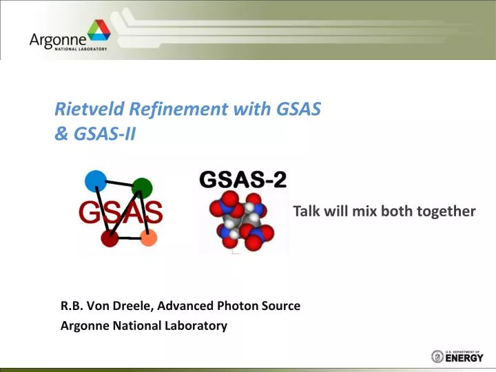
Rietveld Refinement with GSAS & GSAS-II. R.B. Von Dreele , Advanced Photon Source Argonne National Laboratory. Talk will mix both together. What does GSAS do in powder pattern analysis?. Includes: - Rietveld refinement Results Powder pattern plots For publication

E N D
Rietveld Refinement with GSAS & GSAS-II R.B. Von Dreele, Advanced Photon Source Argonne National Laboratory Talk will mix both together
What does GSAS do in powder pattern analysis? • Includes: • - Rietveld refinement • Results • Powder pattern plots • For publication • Bond lengths & angles • Other geometry • CIF (& PDB) files of result • Fourier maps & (some) display • Texture (polefigures) • Utilities • Missing: • Indexing • Structure solution • Must go elsewhere for these. Thanks to Lynn McCusker for maze
Form of GSAS PC-GSAS – thin wrapper GUI forplot expedt genles Keyboard interface only powplot fourier .EXP file, etc. disagl GSAS programs – each is a Fortran exe (common library of routines)
Form of GSAS & EXPGUI widplt forplot expgui expedt GUI genles powplot fourier Keyboard & mouse disagl liveplot EXPGUI – incomplete GUI access to GSAS but with extras
GSAS & EXPGUI interfaces EXPEDT data setup option (<?>,D,F,K,L,P,R,S,X) >EXPEDT data setup options: <?> - Type this help listing D - Distance/angle calculation set up F - Fourier calculation set up K n - Delete all but the last n history records L - Least squares refinement set up P - Powder data preparation R - Review data in the experiment file S - Single crystal data preparation X - Exit from EXPEDT GSAS – EXPEDT (and everything else) – text based menus with help, macro building, etc. (1980’s user interface!) EXPGUI: access to GSAS Typical GUI – edit boxes, buttons, pull downs etc. Liveplot – powder pattern display (1990’s user interface)
GSAS-II: A fresh start • Fill in what’s missing from GSAS: • Indexing • Structure solution • Base code – python • Mixed in old GSAS Fortran • Graphics – matplotlib,OpenGL • Modern GUI – wxPython • Math – numpy,scipy • Current: python 2.7 • All platforms: Windows, Max OSX & Linux GSASII – fresh start
GSAS-II – python code model Slow GUI code – wxPython & common project file name.gpx Fast core processing codes (a few fortran routines) Fast code – numpy array routines Python – ideal for this
GSAS-II: A screen shot – 3 frame layout + console Main menu Data tree Submenu Data tabs Data window Drawing tabs Graphics window NB: Dialog box windows will appear wanting a response
Rietveld results - visualization Easy zoom I/s(I) Normal Probability
Complex peak broadening models m-strain surface NB: mm size & mstrain units
Variance-covariance matrix display Useful diagnostic! High V-covV? Forgot a “hold” Highly coupled parms Note “tool tip”
Structure drawing Polyhedra Van der Waals atoms Balls & sticks Thermal ellipsoids All selectable by atom
Rietveld refinement is multiparameter curve fitting (lab CuKa B-B data) • Result from fluoroapatite refinement – powder profile is curve with counting noise & fit is smooth curve • NB: big plot is sqrt(I) Old GSAS example! ) Iobs+ Icalc| Io-Ic| Refl. positions
So how do we get there? • Beginning – model errors misfits to pattern • Can’t just let go all parameters – too far from best model (minimum c2) False minimum Least-squares cycles c2 True minimum – “global” minimum parameter c2 surface shape depends on parameter suite
Fluoroapatite start – add model (1stchoose lattice & space group) • important – reflection marks match peaks • Bad start otherwise – adjust lattice parameters (wrong space group?)
2nd add atoms & do default initial refinement – scale & background • Notice shape of difference curve – position/shape/intensity errors
Errors & parameters? • position – lattice parameters, zero point (not common) - other systematic effects – sample shift/offset • shape – profile coefficients (GU, GV, GW, LX, LY, etc. in GSAS) • intensity – crystal structure (atom positions & thermal parameters) - other systematic effects (absorption/extinction/preferred orientation) NB – get linear combination of all the above NB2 – trend with 2Q (or TOF) important peak shift wrong intensity too sharp LX - too small a – too small Ca2(x) – too small
Difference curve – what to do next? • Dominant error – peak shapes? Too sharp? • Refine profile parameters next (maybe include lattice parameters) • NB - EACH CASE IS DIFFERENT • Characteristic “up-down-up” • profile error NB – can be “down-up-down” for too “fat” profile
Result – much improved! • maybe intensity differences remain • – refine coordinates & thermal parms.
Result – essentially unchanged Ca F PO4 • Thus, major error in this initial model – peak shapes
Pawley/Rietveld refinement Residual: Exact overlaps - symmetry Io Incomplete overlaps SIc Processing: GSAS – point by point GSAS-II – reflection by reflection Ic Minimize
Least Squares Theory Minimize This is done by setting the derivative of MR to zero ai - initial values of pi Dpi = pi - ai (shift) Normal equations - one for each Dpi; outer sum over observations Solve for Dpi - shifts of parameters, NOT values Matrix form: Ax=v & B = A-1 so x = Bv = Dp
Least Squares Theory - continued Matrix equation Ax=v Solve x = A-1v = Bv; B = A-1 This gives set of Dpi to apply to “old” set of ai repeat until all xi~0 (i.e. no more shifts) Quality of fit – “c2” = M/(N-P) 1 if weights “correct” & model without systematic errors (very rarely achieved) Bii = s2i – “standard uncertainty” (“variance”) in Dpi (usually scaled by c2) Bij/(Bii*Bjj) – “covariance” between Dpi & Dpj Rietveld refinement - this process applied to powder profiles Gcalc - model function for the powder profile (Y elsewhere)
Rietveld Model: Yc = Io{SkhF2hmhLhP(Dh) + Ib} Least-squares: minimize M=Sw(Yo-Yc)2 Io - incident intensity - variable for fixed 2Q kh - scale factor for particular phase F2h - structure factor for particular reflection mh - reflection multiplicity Lh - correction factors on intensity - texture, etc. P(Dh) - peak shape function - strain & microstrain, etc. Ib - background contribution
Peak shape functions – can get exotic! Convolution of contributing functions Instrumental effects Source Geometric aberrations Sample effects Particle size - crystallite size Microstrain - nonidentical unit cell sizes
Gaussian – usual instrument contribution is “mostly” Gaussian CW Peak Shape Functions – basically 2 parts: Lorentzian – usual sample broadening contribution G- full width at half maximum – expression from soller slit sizes and monochromator angle & sample broadening D- displacement from peak position Convolution – Voigt; linear combination - pseudoVoigt
CW Profile Function in GSAS & GSAS-II Thompson, Cox & Hastings (with modifications) Pseudo-Voigt Mixing coefficient FWHM parameter Where Lorentzian FWHM = g and Gaussian FWHM = G
CW Axial Broadening Function Finger, Cox & Jephcoat based on van Laar & Yelon Debye-Scherrer cone 2Q Scan H Slit 2QBragg 2Qmin 2Qi Depend on slit & sample “heights” wrtdiffr. radius H/L & S/L - parameters in function (combined as S/L+H/L; S = H) (typically 0.002 - 0.020) Ä Pseudo-Voigt (TCH) = profile function
How good is this function? Protein Rietveld refinement - Very low angle fit 1.0-4.0° peaks - strong asymmetry “perfect” fit to shape
Bragg-Brentano Diffractometer – “parafocusing” Focusing circle X-ray source Diffractometer circle Receiving slit Incident beam slit Sample displaced Sample transparency Beam footprint Divergent beam optics
CW Function Coefficients – GSAS & GSAS-II Shifted difference Sample shift Sample transparency Gaussian profile Lorentzian profile (plus anisotropic broadening terms) Intrepretation? NB: P term not in GSAS-II; sample shift, meffrefined directly as parameters
b* a* Crystallite Size Broadening Dd*=constant Lorentzian term - usual K - Scherrer const. Gaussian term - rare particles same size? NB: In GSAS-II size is refined directly in mm
b* a* Microstrain Broadening Lorentzian term - usual effect Gaussian term - theory? (No, only a misreading) Remove instrumental part NB: In GSAS-II mstrain refined directly; no conversion needed)
Microstrain broadening – physical model Model – elastic deformation of crystallites Stephens, P.W. (1999). J. Appl. Cryst. 32, 281-289. Also see Popa, N. (1998). J. Appl. Cryst. 31, 176-180. d-spacing expression Broadening – variance in Mhkl
Microstrain broadening - continued Terms in variance Substitute – note similar terms in matrix – collect terms
Microstrain broadening - continued Broadening – as variance 3 collected terms General expression – triclinic – 15 terms Symmetry effects – e.g. monoclinic (b unique) – 9 terms Cubic – m3m – 2 terms
Example - unusual line broadening effects in Na parahydroxybenzoate Sharp lines Broad lines Directional dependence - Lattice defects? Seeming inconsistency in line broadening - hkl dependent
H-atom location in Na parahydroxybenzoate Good DF map allowed by better fit to pattern DF contour map H-atom location from x-ray powder data
Macroscopic Strain Part of peak shape function #5 – TOF & CW d-spacing expression; aij from recip. metric tensor Elastic strain – symmetry restricted lattice distortion TOF: ΔT = (d11h2+d22k2+d33l2+d12hk+d13hl+d23kl)d3 CW: ΔT = (d11h2+d22k2+d33l2+d12hk+d13hl+d23kl)d2tanQ Why? Multiple data sets under different conditions (T,P, x, etc.) NB: In GSAS-II generally available (CW only at present)
Symmetry & macrostrain dij – restricted by symmetry e.g. for cubic DT = d11h2d3for TOF (in GSAS) Result: change in lattice parameters via change in metric coeff. aij’ = aij-2dij/C for TOF aij’ = aij-(p/9000)dij for CW Use new aij’ to get lattice parameters e.g. for cubic
Nonstructural Features Affect the integrated peak intensity and not peak shape Bragg Intensity Corrections: Lh Extinction Absorption & Surface Roughness Preferred Orientation/Texture Other Geometric Factors } diagnostic: Uiso too small!
1 E = 1 + x b 2 3 x x 5 x E = 1 - + - . . . x < 1 2 4 4 8 l 2 1 3 é ù E = 1 - - . . . x > 1 p x ê ú 8 x 2 l 1 2 8 x ë û 2 2 E = E s i n Q + E c o s Q h b l Extinction – only GSAS for now Sabine model - Darwin, Zachariasen & Hamilton Bragg component - reflection Laue component - transmission Combination of two parts
80% 60% 40% 20% 0% 0.0 25.0 50.0 75.0 100.0 125.0 150.0 Sabine Extinction Coefficient Crystallite grain size = Increasing wavelength (1-5 Å) Eh 2Q
What is texture? Nonrandom crystallite grain orientations Random powder - all crystallite orientations equally probable - flat pole figure Pole figure - stereographic projection of a crystal axis down some sample direction Loose powder (100) random texture (100) wire texture Crystallites oriented along wire axis - pole figure peaked in center and at the rim (100’s are 90apart) Orientation Distribution Function - probability function for texture Metal wire
Texture - measurement by diffraction (220) Non-random crystallite orientations in sample (200) Incident beam x-rays or neutrons Sample (111) • Debye-Scherrer cones • uneven intensity due to texture • also different pattern of unevenness for different hkl’s • Intensity pattern changes as sample is turned
Preferred Orientation - March/Dollase Model Uniaxial packing Ellipsoidal Distribution - assumed cylindrical Ro - ratio of ellipsoid axes = 1.0 for no preferred orientation Ellipsoidal particles Spherical Distribution Integral about distribution - modify multiplicity
Texture effect on reflection intensity – Sph. Harm. model • Projection of orientation distribution function for chosen reflection (h) and sample direction (y) • K - symmetrized spherical harmonics - account for sample & crystal symmetry • “Pole figure” - variation of single reflection intensity as fxn. of sample orientation - fixed h • “Inverse pole figure” - modification of all reflection intensities by sample texture - fixed y - Ideally suited for neutron TOF diffraction • Rietveld refinement of coefficients, Clmn, and 3 orientation angles - sample alignment • NB: In GSAS-II as correction & texture analysis
Absorption X-rays - independent of 2Q - flat sample – surface roughness effect - microabsorption effects - but can change peak shape and shift their positions if small (thick sample) Neutrons - depend on 2Q and l but much smaller effect - includes multiple scattering much bigger effect - assume cylindrical sample Debye-Scherrer geometry Diagnostic: thermal parms. too small!
Model - A.W. Hewat For cylinders and weak absorption only i.e. neutrons - most needed for TOF data not for CW data – fails for mR>1 GSAS & GSAS-II – New more elaborate model by Lobanov & alte de Viega – works to mR>10 Other corrections - simple transmission & flat plate (GSAS only for now)
Surface Roughness – Bragg-Brentano & GSAS only Low angle – less penetration (scatter in less dense material) - less intensity High angle – more penetration (go thru surface roughness) - more dense material; more intensity Nonuniform sample density with depth from surface Most prevalent with strong sample absorption If uncorrected - atom temperature factors too small Suortti model Pitschke, et al. model (a bit more stable)