Download
1 / 25
260 likes | 466 Views
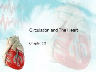
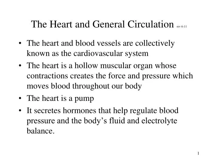
The Heart and General Circulation rev 6-11. The heart and blood vessels are collectively known as the cardiovascular system The heart is a hollow muscular organ whose contractions creates the force and pressure which moves blood throughout our body The heart is a pump

E N D
The Heart and General Circulation rev 6-11 • The heart and blood vessels are collectively known as the cardiovascular system • The heart is a hollow muscular organ whose contractions creates the force and pressure which moves blood throughout our body • The heart is a pump • It secretes hormones that help regulate blood pressure and the body’s fluid and electrolyte balance.
Lab 9-The Heart and General Circulation • Arteries carry blood away from the heart • Veins carry blood to the heart • Arteries and veins are connected to each other by capillaries • The right side of the heart receives returning blood that is low in oxygen (deoxygenated) • Blood moves from the right side of the heart to the lungs where it becomes oxygenated. • Blood is returned to the left side of the heart which circulates it throughout the body
Lab 9-The Heart and General Circulation Anatomy of an Artery, Vein and Capillary • Both arteries and veins have an inner layer of simple, squamous epithelium cells, a middle layer of smooth muscle, and an outer connective layer. • Arteries have thicker muscular layers than veins because they must be able to withstand the high pressures generated by the heart. • Veins are thinner walled and have valves to help keep the blood moving, against gravity, to the heart.
Lab 9-The Heart and General Circulation • Vein valves prevent the backflow of blood. • The smallest arteries are called arterioles; the smallest veins are called venules. These connect via capillaries. • All blood vessels except capillaries have smooth muscles in their walls. • Capillaries are a thin layer of squamous cells which allows for exchange of nutrients, wastes and gases.
Lab 9-The Heart and General Circulation • Layers of an artery • innermost layer is the endothelium • is a layer of flattened, squamous epithelial cells which fit so closely together that they create a slick surface so that blood can flow smoothly. • middle layer which is primarily smooth muscle with elastic connective tissue • outermost layer is a supporting layer of connective tissue which anchors vessels to surrounding tissues
Lab 9-The Heart and General Circulation • Veins • like the walls of arteries, the walls of veins consist of 3 layers of tissue. • Outer 2 layers are much thinner than those of arteries • veins have larger diameters than arteries • the pressure in veins is much lower than that in arteries which is why their walls are not as strong as arteries
Lab 9-The Heart and General Circulation • veins can act as a blood volume reservoir • the larger diameter of veins allows them to stretch to accommodate large volumes of blood at low pressures • because veins can stretch, it is more difficult for them to return blood to the heart against the force of gravity • people who spend a lot of time on their feet may get varicose veins because of this
Lab 9-The Heart and General Circulation • Factors which help veins to return blood to heart • contraction of skeletal muscles • as we move and muscles contract and relax, they press against veins and help push blood to the heart • one way valves in the veins
Lab 9-The Heart and General Circulation • the work of the skeletal muscles helps the valves pump blood. This is called a skeletal muscle pump • movements associated with breathing also help pump blood. This is called a respiratory pump and helps to push blood from the abdomen to the chest and to the heart. • when we breathe, there are pressure changes in the thoracic and abdominal cavities • during inhalation, abdominal pressure increases and squeezes abdominal veins
simultaneously, pressure within the thoracic cavity decreases which dilates the thoracic veins and thus propels the blood. The Heart • External anatomy--heart has blood vessels attached to it • the large vessels are the • aorta which transports blood away from the heart to the entire body. • the pulmonary trunk (pulmonary vein and artery) which transports blood to and from the lungs.
Lab 9-The Heart and General Circulation • The superior and inferior vena cava which return blood to the right atrium • the 4 chambers of the heart are the • 2 atria: the right atrium and the left atrium • 2 ventricles: the right and left ventricles • blood enters the heart through the right atrium, flows through an atrioventricular valve into the right ventricle
Lab 9-The Heart and General Circulation • From the right ventricle, to a pulmonary valve to the pulmonary artery to the lungs • oxygenated blood returns to the heart through the pulmonary vein • and enters the left atrium • flows through another atrioventricular valve to the left ventricle • then through another valve to the aorta to the entire body
Lab 9-The Heart and General Circulation • the heart muscle has its own supply of blood vessels: coronary arteries and coronary veins (see pictures on page 119 of lab manual) • All of the chambers of the heart fill at the same time • note that the pulmonary circulation has the oxygen level of the arteries and veins reversed
Lab 9-The Heart and General Circulation • the heart pumps 2 “circuits” at the same time-- • the right ventricle to the pulmonary trunk • and the left ventricle to the entire body • the outer wall of the left ventricle is thicker than the right ventricle • this ventricle does more work than any other chamber--it must overcome the pressure of the aorta in order to pump blood into it
REMINDER • Look at the bulleted lists on lab manual pages 120 and 121 • you will need to know where these items are located for the lab test • Use the models and charts to help you locate these; don’t try to find them in the fetal pig
Lab 9-The Heart and General Circulation In general, blood vessels are named for the body cavity which they pass through • The aorta modifies its name as it enters different body areas • aortic arch as it leaves heart • thoracic aorta: where aorta straightens out • abdominal aorta: when aorta goes through the diaphragm • after this, the aorta splits to go into each leg; it now becomes the femoral artery
Lab 9-The Heart and General Circulation • Main arterial branches of the aorta (see page 126) • brachiocephalic (first branch off aorta) artery • carotid artery • subclavian (underneath clavicle) artery • renal artery (kidney) • external/internal iliac(ileum=hip bones) arteries • femoral artery
Lab 9-The Heart and General Circulation • Major veins(see page 127) • superior vena cava • inferior vena cava (is the major vein in thoracic, pelvic, and abdominal cavities) • jugular vein
REMINDER • Pathway of circulation; you need to know this • follow instructions on page 121 • Do the sounds of the heart and the heart rate activities (page 121)
Lab 9-The Heart and General Circulation Functional Anatomy • Contraction of the heart muscle is called systole • during systole, blood is pumped out of, or ejected from, the heart into the circulatory system • Relaxation of the heart muscle is called diastole • during diastole, the heart fills with blood
Lab 9-The Heart and General Circulation • Blood Pressure • When blood pressure is taken, • the higher number is the systolic pressure • the lower number is the diastolic pressure • You will be taking each other’s blood pressure with an automatic blood pressure cuff • wrap the cuff snugly around your partner’s arm(with the velcro side facing you, not your partner) • the cuff is placed around the upper part of the arm so the bottom edge rests immediately above the elbow
Lab 9-The Heart and General CirculationREMINDER page 1 of 3: 1.Use the sheep heart and the models to view internal anatomy. Learn all the heart structures on the models. Remember to label figure 13.5 to include the heart chambers--atria and ventricles. 2. Locate all blood vessels from the bulleted lists on pages 120-121 on the models. Use the figures on lab manual pages 124-125 to help you. 3. See the instructions on page 121 for the pathway of circulation and follow this activity.
REMINDER page 2 of 3: 4. Do the sounds of the heart and the heart rate activities 5. Perform the blood pressure activities on pages 124-125. • We will also begin dissecting the fetal pig today. Remember that everyone needs to participate in the dissection. • Tie long hair back so it doesn’t mix with the inside of the pig when you bend down to look at it.
REMINDER page 2 of 3: 7. You need to locate the following endocrine glands: thyroid, thymus, and pancreas (see diagram on page 104 of the lab manual). • DO NOT locate the adrenal glands; they are very hard to find. • Remember to use Table 11.1 to help you learn about these glands, their hormones and the function of these hormones.