Download
1 / 82
820 likes | 963 Views
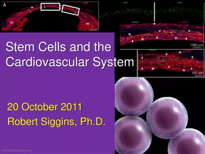
Stem Cells and the Cardiovascular System. 20 October 2011 Robert Siggins, Ph.D. stemcellschool.com. Outline and Objectives. What are stem cells (SCs)? Which SCs contribute to the adult CV system? What are the physiological and pathophysiological roles for SCs in the CV system?.

E N D
Stem Cells and the Cardiovascular System 20 October 2011 Robert Siggins, Ph.D. stemcellschool.com
Outline and Objectives • What are stem cells (SCs)? • Which SCs contribute to the adult CV system? • What are the physiological and pathophysiological roles for SCs in the CV system?
Stem Cell “Theory” • 1855 First eluded to by (Remak) Virchow Omnis cellula e cellula • RediOmne vivum ex ovo • 1875 ”Embryonic rest” proposed by Cohnheim (cancer stem cells) • 1917 HSC proposed by Pappenheim • 1961 McCulloch and Till describe CFU-s
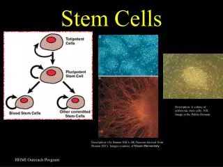
Definition of SCs • Totipotent, pluripotent, or multipotent • Self-renewal • Clonogenic • Niche http://icanhascheezburger.com
Totipotent = ability to give rise to all cells of the body and all cells that constitute the extraembryonic tissues (i.e. placenta) Self-renewal Pluripotent = ability to make all cells of the body (all three germ layers) Multipotent = ability to differentiate into more than one cell type (usually germ layer restricted) autismpedia.org
Embryonic SCs (ESCs) are pluripotent Tissue-restricted SCs are Multipotent! stemcellresources.org
UEA-1 (ulexeuropaeus agglutinin-1) or SSEA-4 (stage-specific embryonic antigen-4) eliminates teratoma-initiating cells! Physiol Rev 85:1373-1416, 2005
Definition of SCs • Totipotent, pluripotent, or multipotent • Self-renewal • Clonogenic • Niche
Symmetric and Asymmetric Divisions http://xarquon.jcu.cz/edu/hematologie/03kmenove_bunky/03stem_lineages.htm
Cell Extrinsic and Intrinsic Models Regulated by niche attachment Regulated by niche cell polarity Regulated by intrinsic polarity
Definition of SCs • Totipotent, pluripotent, or multipotent • Self-renewal • Clonogenic • Niche
Cardiac Stem Cells (CSCs) Large clone of c-kit+ cells (green) generated by a single rat c-kit+ CSC. Physiol Rev 85:1373-1416, 2005
Cardiac Stem Cells (CSCs) Clonogenic cells in differentiating medium acquire the myocyte phenotype (α-sarcomeric actin, red). Physiol Rev 85:1373-1416, 2005
Cardiac Stem Cells (CSCs) Clonogenic cells acquire the SMC phenotype (α-smooth muscle actin, magenta). Physiol Rev 85:1373-1416, 2005
Cardiac Stem Cells (CSCs) Clonogenic cells acquire the EC phenotype (vWF, yellow). Physiol Rev 85:1373-1416, 2005
Definition of SCs • Totipotent, pluripotent, or multipotent • Self-renewal • Clonogenic • Niche
Niches are specialized microenvironmnets comprising support cells, soluble factors, ECM, SCs, and progenitor cells Every niche is unique!
Cardiac niches and putative supporting cells. Apical (A, B, and F) and atrial (C–E) niches contain CSCs and LCCs. (A) c-kit+ (green) Ets1+ (white; EC progenitors). (B) MDR1+ cells (white) GATA-4+ (magenta dots); α-sarcomeric actin+GATA-4+ (red; myocyte precursors). GATA-4+ cardiac progenitors (arrowheads). (C) Sca-1+MEF2C+ (yellow & white dots; myocyte progenitors); Sca-1+von Willebrand factor+ (green; EC precursor). (D–F) Connexin 43 and E- and N-cadherin in niches containing c-kit+ (D & F, green) and Sca-1+ (E, yellow) CSCs and LCCs. GATA-4+ (D, magenta dots) and MEF2C+ (E & F, yellow dots). Connexin 43 (yellow dots), E-cadherin (green dots), and N-cadherin (white dots) Nuclei are stained by propidium iodide (blue). PNAS 103:9226-9231, 2006
Cardiac niches and putative supporting cells. (G–J) Connexin 43 and 45 and N- and E-cadherin in atrial niches containing c-kit CSCs–LCCs (green) are represented by yellow dots located between two CSCs–LCCs (arrowheads), a CSC–LCC and a fibroblast (arrows), and a CSC–LCC and a myocyte (double arrowheads); see Insets. Nuclei are stained by propidium iodide (blue). PNAS 103:9226-9231, 2006
Formation of Gap Junctions PNAS 103:9226-9231, 2006
PNAS 103:9226-9231, 2006 Myocyte turnover and life span (A and B) Apical section illustrating bright (arrows), intermediate (open arrowheads), and dim (arrowheads) BrdU+ myocyte nuclei (yellow) after 10 weeks of chasing. Myocytes are stained by cardiac myosin (B, red).
Myocyte Turnover (C) % of BrdU-bright, -int, and -dim myocyte nuclei. (Right) Bars document the total number of BrdU+ myocytes and total number of myocytes in the atria, base–mid region, and apex. PNAS 103:9226-9231, 2006
Myocyte Turnover PNAS 103:9226-9231, 2006
Myocyte Turnover PNAS 103:9226-9231, 2006
Myocyte Lifespan PNAS 103:9226-9231, 2006
ESCs versus Adult SCs (ASCs) • ESCs • Embryolically derived • Not tissue-specific • Pluripotent • Requires in vitro fertilized egg for derivation (ethical concerns) • ASCs • Tissue resident • Tissue-specific • Multipotent • No ethical problems with their clinical application
Outline and Objectives • What are stem cells (SCs)? • Which SCs contribute to the adult CV system? • What are the physiological and pathophysiological roles for SCs in the CV system?
A B C D Identification of Mitotic Spindles in Dividing Myocytes from Infarcted Hearts (A) blue = organization of tubulin in the mitotic spindle (arrows) (B) green = metaphase (arrowheads) (C) green and blue = combination of tubulin and metaphase chromosomes (arrows and arrowheads) (D) red = sarcomeric α-actin, blue = tubulin, and green = chromosomes in metaphase (arrows and arrowheads) N Engl J Med 344:1750-1757, 2001
A Myocyte in the Process of Cytokinesis Arrows = actin Red = staining of myocyte sarcomeric α-actin green = PI labeling of chromosomes N Engl J Med 344:1750-1757, 2001
Effects of MI on the Number of Mitotic Myocytes N Engl J Med 344:1750-1757, 2001
The Y Chromosome in Transplanted Hearts. • Myocytes (A and B) • Smooth-muscle cells (C and D), Endothelial cells in both coronary arterioles (E and F), and capillary endothelial cells (G and H) • Blue areas = PI staining in nuclei, Green areas indicate Y chromosome • The red areas indicate sarcomeric α-actin in B, of smooth-muscle α-actin in D, and of factor VIII in F and H. N Engl J Med 346:5-15, 2002
The Y Chromosome in Transplanted Hearts. In I, J, and K, the bright blue, fluorescent areas and the arrows indicate the presence of Ki-67, and the yellow areas show the Y chromosomes. N Engl J Med 346:5-15, 2002
ASCs of the CV system • Isolated based on functional assessments or surface phenotypes • Cardiac Stem Cells (CSCs) and Endothelial Progenitor Cells (EPCs) • Possible Vascular Progenitor Cell (VPC); resident in vessel walls
Surface Markers for EPCs • Mouse • VEGFR2, LinNeg, c-kit, Sca-1, VE-cadherin • Human • CD133, VEGFR2, VE-cadherin, CD34
Vasculogenesis • Aggregation of de-novo-forming angioblasts into a primitive vascular plexus • Hemangioblast Flk1+ cells produce ECs and hematopoietic cells • Earliest marker of angioblast precursors Flk1/VEGFR-2 • Complex remodeling processgrowth, migration, sprouting and pruning lead to development of functional circulatory system