Download
1 / 44
440 likes | 523 Views
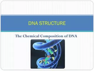
DNA STRUCTURE. NUCLEIC ACIDS. Nucleic acids are polymers Monomer---nucleotides Nitrogenous bases Purines Pyrimidines Sugar Ribose Deoxyribose Phosphates +nucleoside=nucleotide. }. Nucleosides. The Sugars. PYRIMIDINES. PURINES. The Bases. Bases of DNA (and RNA). Purines:.

E N D
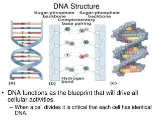
NUCLEIC ACIDS • Nucleic acids are polymers • Monomer---nucleotides • Nitrogenous bases • Purines • Pyrimidines • Sugar • Ribose • Deoxyribose • Phosphates • +nucleoside=nucleotide } Nucleosides
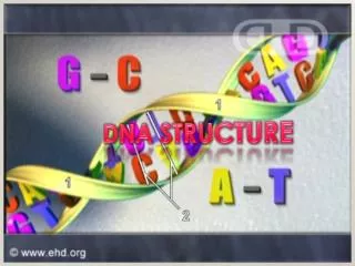
PYRIMIDINES PURINES The Bases
Bases of DNA (and RNA) Purines: Pyrimidines: DNA only RNA only
Resume Chemical Structure of DNA and RNA Figure 4.1 The C is named 1’-5’ 1’ 4’ 2’ Nucleotide Nucleoside DNA RNA
Nucleotides and Nucleosides Nucleotides are nucleosides + phosphate
OH P O CH2 Base HO O C C H H O C C H H H H or OH Nucleic Acids make up 13-34% of the dry weight in bacteria deoxyribonucleic acid (DNA) and ribonucleic acid (RNA) Nucleoside: base + sugar Nucleotide: a building block • Sugar: • RNA – ribose (OH) • DNA – deoxyribose (H) • Bases: • adenine (A), cytosine (C), guanine (G), • thymine (T) • RNA uses uracil (U) instead of thymine • certain nucleotides serve as a storage of energy and reducing power • e.g. ATP -> ADP -> AMP hydrolysis (energy is released)
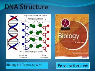
Advantages to Double Helix • Stability---protects bases from attack by H2O soluble compounds and H2O itself. • Provides easy mechanism for replication
Physical Structure (cont’d) • Chains are anti-parallel (i.e in opposite directions) • Diameter and periodicity are consistent • 2.0 nm • 10 bases/ turn • 3.4 nm/ turn • Width consistent because of pyrimidine/purine pairing
G-C Content • A=T, G=C, but AT≠GC • Generally GC~50%, but extremely variable • EX. • Slime mold~22% • Mycobacterium~73% • Distribution of GC is not uniform in genomes
CONSEQUENCES OF GC CONTENT • GC slightly denser • Higher GC DNA moves further in a gradient • Higher # of base pairs=more stable DNA, i.e. the strands don’t separate as easily.
Cruciform Structures Another adaptation to supercoiling Associated with palindromes
DNA is Dynamic • Like proteins, DNA has 3º structure • Why so many deviations from normal conformation? • Effects on transcription (gene expression) • Enhances responsiveness • May also serve in packaging • NOTE: most cellular DNA exists as protein containing supercoils
Denaturation of DNA • Denaturation by heating. • How observed? • A260 • For dsDNA, A260=1.0 for 50 µg/ml • For ssDNA and RNA A260=1.0 for 38 µg/ml • For ss oligos A260=1.0 for 33 µg/ml • Hyperchromic shift The T at which ½ the DNA sample is denatured is called the melting temperature (Tm)
Importance of Tm • Critical importance in any technique that relies on complementary base pairing • Designing PCR primers • Southern blots • Northern blots • Colony hybridization
Factors Affecting Tm • G-C content of sample • Presence of intercalating agents (anything that disrupts H-bonds or base stacking) • Salt concentration • pH • Length
Renaturation • Strands can be induced to renature (anneal) under proper conditions. Factors to consider: • Temperature • Salt concentration • DNA concentration • Time
What Do Cot Curves Reveal? • Complexity of DNA sample • Reveals important info about the physical structure of DNA • Can be used to determine Tm for techniques that complementary base pairing.
Complexity of DNA- FactorsRepetitive Sequences • Single Copy Genes • Highly repetitive (hundreds to millions) • Randomly dispersed or in tandem repeats • Satellite DNA • Microsatellite repeats • Miniisatellite repeats • Middle repetitive (10- hundreds) • Clustered • Dispersed • Slightly repetitive (2-10 copies)
Renaturation curves of E. coli and calf DNA Highly repetitive sequences Middle repetitivesequences Unique sequences
RNA • Types • mRNA • tRNA • rRNA • It’s still an RNA world • snRNA • siRNA • Ribozymes
Behavior in Acids • Dilute or mild acidic conditions • Intermediate conditions. EX. 1N HCl @ 100ºC for 15m : Depurination • Harsher treatment-EX. 2-6N HCl, higher temps: Depyrimidination. • NOTE: some phosphodiester bond cleavage observed during depurination, much more during depyrimidination
Behavior in Bases • N-glycosidic bonds stable in mild alkaline conditions • DNA melts • Phosphodiester linkages in DNA and RNA show very different behavior in weak bases (EX 0.3 N KOH @37ºC ~1 hr.)
RNA Hydrolysis in Alkaline Solutions 2,3 cyclic nucleotide
Hydrolysis by Enzymes • Nuclease—catalyzes hydrolysis of phosphodiester backbone • Exonucleases • Endonucleases • General. Ex DNAse I • Specific Ex. Restriction endonucleases • Ribozymes
RIBOZYMES • Catalytic RNA • Can work alone or with proteins • Therapeutic applications?
SEQUENCING • Purpose—determine nucleotide sequence of DNA • Two main methods • Maxam & Gilbert, using chemical sequencing • Sanger, using dideoxynucleotides
The Sanger Technique • Uses dideoxynucleotides (dideoxyadenine, dideoxyguanine, etc) • These are molecules that resemble normal nucleotides but lack the normal -OH group.
Because they lack the -OH (which allows nucleotides to join a growing DNA strand), replication stops. Normally, this would be where another phosphate Is attached, but with no -OH group, a bond can not form and replication stops
The Sanger Method Requires • Multiple copies of single stranded template DNA • A suitable primer (a small piece of DNA that can pair with the template DNA to act as a starting point for replication) • DNA polymerase (an enzyme that copies DNA, adding new nucleotides to the 3’ end of the template • A ‘pool’ of normal nucleotides • A small proportion of dideoxynucleotides labeled in some way ( radioactively or with fluorescent dyes)
The template DNA pieces are replicated, incorporating normal nucleotides, but occasionally and at random dideoxy (DD) nucleotides are taken up. • This stops replication on that piece of DNA • The result is a mix of DNA lengths, each ending with a particular labeled DDnucleotide. • Because the different lengths ‘travel’ at different rates during electrophoresis, their order can be determined.