Download
1 / 93
1.07k likes | 1.55k Views
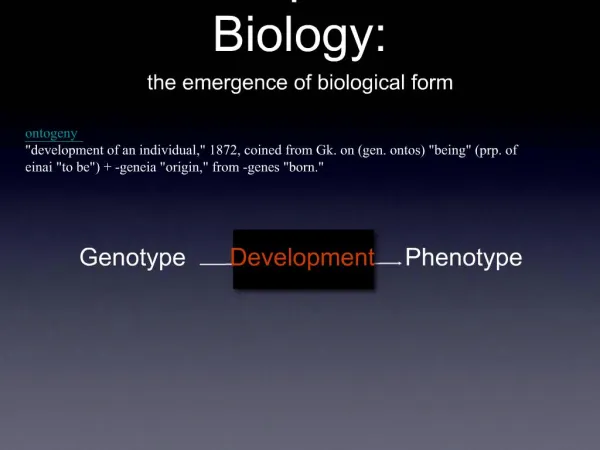
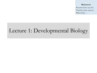
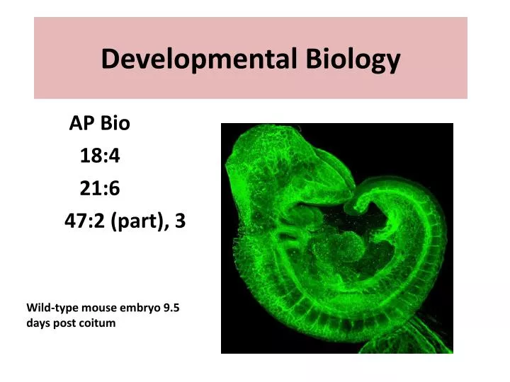
Developmental Biology. AP Bio 18:4 21:6 47:2 (part), 3. Wild-type mouse embryo 9.5 days post coitum. Our focus:. Timing and Coordination Gene Expression Interactions, Cell Signaling. Insights into development have been obtained froms studying. Slime Molds Nematode worm C. elegans

E N D
Developmental Biology AP Bio 18:4 21:6 47:2 (part), 3 Wild-type mouse embryo 9.5 days post coitum
Our focus: Timing and Coordination Gene Expression Interactions, Cell Signaling
Insights into development have been obtained froms studying • Slime Molds • Nematode worm C. elegans • Fruit Flies • Zebrafish • Frog Embryos • Chick Embryos • Mice
Fig. 18-14 (a) Fertilized eggs of a frog (b) Newly hatched tadpole
A program of differential gene expression leads to the different cell types in a multicellular organism • During embryonic development, a fertilized egg gives rise to many different cell types • Cell types are organized successively into tissues, organs, organ systems, and the whole organism • Gene expression orchestrates the developmental programs of animals
A Genetic Program for Embryonic Development • The transformation from zygote to adult results from cell division, cell differentiation, and morphogenesis
Cell differentiation is the process by which cells become specialized in structure and function • The physical processes that give an organism its shape constitute morphogenesis • Differential gene expression results from genes being regulated differently in each cell type • Materials in the egg can set up gene regulation that is carried out as cells divide
Cytoplasmic Determinants and Inductive Signals • An egg’s cytoplasm contains RNA, proteins, and other substances that are distributed unevenly in the unfertilized egg • Cytoplasmic determinants are maternal substances in the egg that influence early development • As the zygote divides by mitosis, cells contain different cytoplasmic determinants, which lead to different gene expression
Fig. 18-15a Unfertilized egg cell Sperm Nucleus Fertilization Two different cytoplasmic determinants Zygote Mitotic cell division Two-celled embryo (a) Cytoplasmic determinants in the egg
The other important source of developmental information is the environment around the cell, especially signals from nearby embryonic cells • In the process called induction, signal molecules from embryonic cells cause transcriptional changes in nearby target cells • Thus, interactions between cells induce differentiation of specialized cell types
Work with the nematode C. elegans has shown that induction requires the transcriptional regulation of genes in a particular sequence.
Fig. 18-15b NUCLEUS Early embryo (32 cells) Signal transduction pathway Signal receptor Signal molecule (inducer) (b) Induction by nearby cells
Sequential Regulation of Gene Expression During Cellular Differentiation • Determination commits a cell to its final fate: it is the progressive restriction of developmental potential as the embryo develops • Determination precedes differentiation • Cell differentiation is marked by the expression of tissue-specific proteins
Myoblastsproduce muscle-specific proteins and form skeletal muscle cells MyoDis one of several “master regulatory genes” that produce proteins that commit the cell to becoming skeletal muscle The MyoD protein is a transcription factor that binds to enhancers of various target genes For example
Fig. 18-16-1 Nucleus Master regulatory gene myoD Other muscle-specific genes DNA Embryonic precursor cell OFF OFF
Fig. 18-16-2 Nucleus Master regulatory gene myoD Other muscle-specific genes DNA Embryonic precursor cell OFF OFF OFF mRNA MyoD protein (transcription factor) Myoblast (determined)
Fig. 18-16-3 Nucleus Master regulatory gene myoD Other muscle-specific genes DNA Embryonic precursor cell OFF OFF OFF mRNA MyoD protein (transcription factor) Myoblast (determined) mRNA mRNA mRNA mRNA Myosin, other muscle proteins, and cell cycle– blocking proteins MyoD Another transcription factor Part of a muscle fiber (fully differentiated cell)
Why doesn’t myoD change any type of embryonic cell? • Probably a combination of regulatory genes are necessary for differentiation is required.
Pattern Formation: Setting Up the Body Plan • Pattern formation is the development of a spatial organization of tissues and organs • In animals, pattern formation begins with the establishment of the major axes • Positional information, the molecular cues (cytoplasmic determinants and inductive signals) control pattern formation, and tell a cell its location relative to the body axes and to neighboring cells
Pattern formation has been extensively studied in the fruit fly Drosophila melanogaster • Combining anatomical, genetic, and biochemical approaches, researchers have discovered developmental principles common to many other species, including humans
The Life Cycle of Drosophila • In Drosophila, cytoplasmic determinants in the unfertilized egg determine the axes before fertilization • After fertilization, the embryo develops into a segmented larva with three larval stages
Fig. 18-17a Thorax Head Abdomen 0.5 mm Dorsal Right BODY AXES Posterior Anterior Left Ventral (a) Adult
Fig. 18-17b Follicle cell Egg cell developing within ovarian follicle 1 Nucleus Egg cell Nurse cell Egg shell Unfertilized egg 2 Depleted nurse cells Fertilization Laying of egg Fertilized egg 3 Embryonic development Segmented embryo 4 0.1 mm Body segments Hatching Larval stage 5 (b) Development from egg to larva
Genetic Analysis of Early Development: Scientific Inquiry • Edward B. Lewis, Christiane Nüsslein-Volhard, and Eric Wieschaus won a Nobel 1995 Prize for decoding pattern formation in Drosophila • Homeotic genes control pattern formation in the late embryo, larva, and adult.
Fig. 18-18 A mutation in regulatory genes, called homeotic genes, caused this. Eye Leg Antenna Wild type Mutant
Homeotic Genes • One example are the Hox and ParaHox genes which are important for segmentation,another example is the MADS-box-containing genes in the ABC model of flower development. Chap 21:6
The Homeobox • Homeotic genes contain a 180 nucleotide sequence called a homeobox found in regulatory genes . • This homeobox has been found in inverts and verts as well as plants.
The homeobox DNA sequence evolved very early in the history of life and has been conserved virtually unchanged for millions of years. Differences arise due to different gene expressions.
Molecular basis of differentiation: • The A, B, and C genes are transcription factors. Different transcription factors are needed together to turn on a developmental gene program--such as A and B needed to initiate the program for petals. What turns on the different transcription factors in different cells? • Induction and inhibition by one cell signaling to a neighboring cell.
Fate Mapping • Fate maps are general territorial diagrams of embryonic development • Classic studies using frogs indicated that cell lineage in germ layers is traceable to blastula cells *Chap 47 (3) http://education-portal.com/academy/lesson/how-fate-mapping-is-used-to-track-cell-development.html
Fig. 47-21 Epidermis Centralnervoussystem Epidermis 64-cell embryos Notochord Blastomeresinjected with dye Mesoderm Endoderm Larvae Neural tube stage(transverse section) Blastula (a) Fate map of a frog embryo (b) Cell lineage analysis in a tunicate
Fig. 47-21a Epidermis Centralnervoussystem Epidermis Notochord Mesoderm Endoderm Neural tube stage(transverse section) Blastula (a) Fate map of a frog embryo
Axis Establishment • Maternal effect genes encode for cytoplasmic determinants that initially establish the axes of the body of Drosophila • These maternal effect genes are also called egg-polarity genes because they control orientation of the egg and consequently the fly
Bicoid: A Morphogen Determining Head Structures • One maternal effect gene, the bicoid gene, affects the front half of the body • An embryo whose mother has a mutant bicoid gene lacks the front half of its body and has duplicate posterior structures at both ends
Fig. 18-19a EXPERIMENT Tail Head A8 T1 T2 A7 T3 A6 A1 A5 A2 A3 A4 Wild-type larva Tail Tail A8 A8 A7 A7 A6 Mutant larva (bicoid)
Fig. 18-19b RESULTS Fertilization, translation of bicoid mRNA Anterior end 100 µm Bicoid mRNA in mature unfertilized egg Bicoid protein in early embryo
Fig. 18-19c CONCLUSION Nurse cells Egg bicoid mRNA Bicoid mRNA in mature unfertilized egg Developing egg Bicoid protein in early embryo
This phenotype suggests that the product of the mother’s bicoid gene is concentrated at the future anterior end • This hypothesis is an example of the gradienthypothesis, in which gradients (amounts) of substances called morphogens establish an embryo’s axes and other features
The bicoid research is important for three reasons: – It identified a specific protein required for some early steps in pattern formation – It increased understanding of the mother’s role in embryo development – It demonstrated a key developmental principle that a gradient of molecules can determine polarity and position in the embryo
Cancer results from genetic changes that affect cell cycle control • The gene regulation systems that go wrong during cancer are the very same systems involved in embryonic development
Types of Genes Associated with Cancer • Cancer can be caused by mutations to genes that regulate cell growth and division • Tumor viruses can cause cancer in animals including humans • Oncogenesare cancer-causing genes • Proto-oncogenes are the corresponding normal cellular genes that are responsible for normal cell growth and division • Conversion of a proto-oncogene to an oncogene can lead to abnormal stimulation of the cell cycle
Fig. 18-20 Proto-oncogene DNA Point mutation: Gene amplification: Translocation or transposition: within the gene within a control element New promoter Oncogene Oncogene Normal growth- stimulating protein in excess Normal growth-stimulating protein in excess Normal growth- stimulating protein in excess Hyperactive or degradation- resistant protein