Download
1 / 31
310 likes | 430 Views
Urogyn Disorders & Oncology Complications of Gyn Procedures. Chapters 111 and 112. Incontinence. Anatomic Structures that provide continence: Detrusor muscle Urethra Urethral sphincter 2 posterior pubourethral ligaments Autonomic nervous system Pudendal nerves.

E N D
Urogyn Disorders & OncologyComplications of Gyn Procedures Chapters 111 and 112
Incontinence • Anatomic Structures that provide continence: • Detrusor muscle • Urethra • Urethral sphincter • 2 posterior pubourethral ligaments • Autonomic nervous system • Pudendal nerves
Classification of Incontinence • Urinary stress incontinence • Increased intra-abdominal pressure • Urge incontinence • Detrusor muscle instability (spasm) • Total incontinence • Usu. Due to urinary fistula • Overflow incontinence • Urinary retention from a hypotonic detrusor muscle (feeling of post-void bladder fullness)
Uterine Prolapse • Caused by the loss of proper alignment when supporting structures fail • 1st degree: cervix remains within the vagina • 2nd degree: cervix protrudes beyond introitus • 3rd degree: entire uterus outside the vulva aka procidentia
Subdivisions of vaginal prolapse • Upper anterior vag. wall: cystocele • Lower anterior vag. wall: urethral displacement • Upper posterior vag. wall: enterocele • Lower posterior vag. wall: rectocele • Inversion of vaginal is known as vault prolapse
Urethral Syndrome • Complex of symptoms including: frequency, urgency, dysuria, suprapubic discomfort, post void fullness, incontinence, and/or dypareunia with NO urologic pathology • Cause not yet determined • Diagnosed by exclusion • No clear cut treatments, may try antibiotics, urethral suppositories or surgery
Gynecologic Malignancies • 13% of cancers in women • It is most important to recognize that a women presenting to the ED with ascites has a gynecologic malignancy until proven otherwise
Ovarian CA • Peak age 55-65 with 1-2% incidence if (-)fhx • Risk factors: infertility, low parity, high fat diet, hx of breast/colon CA, family hx and lactose intolerance • OCP are thought to be protective • Stage1: one/both ovaries • Stage 2: extension to the pelvis • Stage 3: growth outside the pelvis • Stage 4: distant mets
Uterine CA • Most common gyn CA with peak age 58 • Most common type is adenocarcinoma • Risk factors: early menses, late menopause, nulliparity, obesity, DM, HTN, unopposed estrogen use • Most present with postmenopausal bleeding
Uterine CA stages • Stage 1: confined to the uterus corpus • Stage 2: involvement of the cervix • Stage 3: Extends to the uterine serosa, ovary, vagina, and para-aortic/pelvic nodes • Stage 4: Involvement of the bladder/bowel, inguinal/intra-abdominal lymph nodes • Recurrences at vagina, vaginal cuff and pelvis
Cervical CA • Ave age 54 • Risk factors: multiple partners, early coitus, high-risk male partners, smoking, HPV and HIV (considered AIDS defining illness) • Most commonly squamous type • Present with DUB, post-coital bleeding, vag d/c, pain, or leg swelling
Staging of Cervical CA • Stage 1: Strictly cervix • Stage 2: Beyond cervix but not to pelvic sidewall • Stage 2A: No parametrial involvement • Stage 2B: Parametrial involvement • Stage 3: Extends to the pelvic sidewall • Stage 4: Beyond the true pelvis, bladder or rectum involvement
Vaginal CA • 1-2% with mean age of 60-65yrs • Usually squamous type • Risk factors: DES, immunosuppression, low socioeco status, chronic irritation, radiation for other CAs, multiple sex partners • Presents with abnormal vaginal bleeding • Embyonal rhabdomyosarcoma: kids under 5yo with vaginal bldg, d/c or grapelike masses
Vulvar CA • 1-4% of gyn CA, most older than 55 • Usu squamous cell type • May be due to HPV with other risk factors of : smoking, immunodeficiency, exposure to aniline dye (benzene) • Present with mass, pruritus, pain or ulceration
Gestational Trophoblastic Dx • Four types • Hydatiform mole • May be malignant or benign (partial or complete) • Invasive mole • Choriocarcinoma • Placental site tumors
Complications of Gyn Malignancies • Most common is vaginal bleeding • Genital tract bleeding, GI obstruction, masses or ascites, fistulae, obstructive uropathy, mets, lymphadema, and hypercoagulable state (DVT/PE) • Also may have complications due to radiation and chemotherapy
Complications of Gyn procedures • Postop presents to the ED usu with pain, fever, or vaginal bleeding • *don’t forget most fevers within 24hrs is not due to infection but atelectasis, hypersensitivity to antibiotics, pyogenic reaction to tissue trauma & hematoma • Some tenderness is normal, not rebound
Complications of laparoscopy • US rates as low as 0.22% for major complications and 10% for minor • Include thermal injury to the bowel, perforation of a viscous, bleeding or other vascular injury, ureteral or bladder injuries, incisional hernia or wound dehiscence
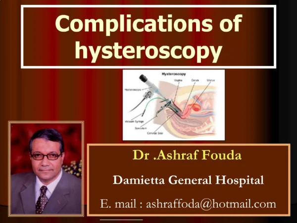
Complications of Hysteroscopy • Fluid overload • Uterine perforation with damage to intra-abdominal organs • Infection • Toxic shock syndrome • Anesthesia reaction • Post-op bleeding • embolism
Wound infection • >90% occur within first two weeks • Initially local cellulitis • May progress to wound dehiscence • Consult the gyn surgeon • Start PCN based antibiotic and aggressive wound care management
Hysterectomy complications • Infected vaginal cuff hematoma, cellulitis, or abscess • Triple antibiotic IV tx and admission • Ampicillin 2g • Gentamycin 1.5mg/kg • Clindamycin 900mg
Ureteral Injury • Three types • Crushing • Transection • Ligation • CT with IV contrast, UA • May have fever and CVA tenderness • Also consider urinary retention from bladder atony or post op pain
Vesicovaginal Fistula • May occur after total abdominal hysterectomy • Present 10-14days post op with watery vaginal d/c • Gyn consult • Usu place a foley catheter
Osteomyelitis Pubis • Rare • Occurs 6-8wks post-op • Tenderness along pubic symphysis esp with ambulation • Have low-grade fever, elevated sed rate and leukocytosis • Admit to IV antibiotics
Wound seroma/hematoma • Hematomas more common in transverse incisions • Open if infection suspected • Ultrasound of wound helpful • Seroma is a collection of serous fluid and may drain spontaneously
Postconization bleeding • Cervical CA or high grade lesions often treated with loop electrocautery or cold-knife conization • Most present with bleeding • Severe hemorrhage may occur and is usually 7 days post-op
Septic Pelvic Thrombophlebitis • Diagnosis of exclusion • 2-4d post-op • Fever, tachycardia, GI distress, unilateral abdominal pain • 50-67% have a palpable cord • Tx with heparin and antibiotics (esp against Bacteroides ie Clindamycin)
Induced Abortion • Termination by: • Instrumental evacuation through the vagina • Stimulation of uterine contraction • Major surgical procedures Early complications (24hrs) bleeding and pain • uterine perforation, retained products or cervical laceration Delayed complications(24hrs-4wks) • Bleeding, endometritis, retained products Late complications • Depression, amenorrhea, Rh isoimmunization
Brachytherapy • Treatment of malignant sources by radioactive implantation • Most used for cervical and uterine CA • Sigmoid, rectosigmoid, and rectum susceptible to injury also the bladder • Problems range from cystitis to tissue necrosis with infection and fistula formation • Hyperbaric O2 has shown to help
Postoperative Fatigue • 60-90% of hysterectomy patients experience profound fatigue • May take up to 10wks before able to return to full normal activities • Consider depression and iron deficiency anemia
Ovarian hyperstimulation syndrome • May be life threatening, 1-2% • Abdominal distention, ovarian enlargement, and weight gain • Severe form: ascites, electrolyte imbalances, pleural effusions, hypovolemia • Increased coagulability and renal failure • Abdominal and pelvic exam is contraindicated due to fragility of ovaries which may lead to rupture/hemorrhage