Download
1 / 29
290 likes | 321 Views
Explore the intricate nervous system of chordates, focusing on the primary brain vesicles, hindbrain divisions, midbrain functions, and forebrain structures. Learn about sensory processing in chordates and brain evolution in vertebrates.

E N D
Comparative Chordate Anatomy: II Nervous System(T.Y.B.Sc.) Dr. Rahul B. Patil, Department of Zoology, Veer Wajekar A.S.C. College, Phunde
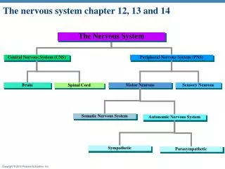
Primary Brain Vesicles • Prosencephalon (Forebrain) • Smell • Mesoncephalon (Midbrain) • Vision • Rhombencephalon (Hindbrain) • Hearing Figure 15.1: Primary brain vesicles
Primary Brain Vesicles (con’t) Figure 15.2: Basic brain plan. Figure 15.3: Brain divisions.
Hindbrain • Myelencephalon • Medulla oblongata • Involuntary reflexes • Vagal lobe • Metencephalon • Cerebellum • Roof of metencephalon • Reflex control of skel. muscle • Pons Figure 15.4: Neural tube development.
Hindbrain (con’t) • Lateral ventricles • Two cerebral hemispheres • Posterior choroid plexus • Roof in hindbrain • 4th ventricle tissue • Cerebral spinal fluid • Tela choroidea • Roof of medulla • Thin membrane Figure 15.5: Choroid plexus shown of larval anuran.
Divisions and Vesicles Figure 15.6: Divisions of the brain and ventricles numbered.
Midbrain • No subdivisions • Optic lobes • Optic reflex centers • Well developed in birds • Auditory lobes • Caudal to optic lobes • Superior (optic) and inferior (auditory) colliculi- when lobes occur together • Corpora quadrigemina collectively Figure 15.7: Mesoncephalon and tectum region.
Midbrain (con’t) • 3rd ventricle • Optic ventricles- extension to optic lobe • Ventricles disappear in higher phylogeny • Cerebral aqueduct • Restricted passageways • Conducts 3rd and 4th ventricle • Aqueduct of Sylvius when restricted further Figure 15.8: Cerebral aqueduct and ventricles of brain.
Forebrain- Diencephalon • Optic chiasma • Two optic nerves cross • Pituitary gland • Caudal to optic chiasma • Saccusvasculosus • Posterior to pituitary in some fish • Depth receptor Figure 15.9: Regions of the diencephalon of a shark with third ventricle in red
Forebrain- Diencephalon (con’t) • Hypothalamus • Floor of diencephalon • Autonomic nervous system • Thalamus • Walls of diencephalon • 3rd ventricle cavity • Communicates with lateral ventricles • Foramen of Monro Figure 15.10: Medial view of the brain showing thalamus and hypothalamus of the diencephalon.
Forebrain- Diencephalon (con’t) • Epithalamus • Several evaginations • Roof of diencephalon • Paraphysis anteriorly • Epiphyseal complex • Pineal • Photoreceptors • Parapineal • Pineal eye (3rd eye) Figure 15.11: Epithalamus; gross mid-sagittal section of the human brain. Figure 15.12: Pineal in detail
Forebrain- Telencephalon • Cerebral hemispheres posterior • Rhinencephalon anterior • Olfaction • Lower vertebrates • Rhinencephalon prominent • Hemispheres smaller • Higher vertebrates • Hemispheres increase in size • Olfactory get smaller Figure 15.13: Front section of cerebral hemisphere formation
Craniate Brains Figure 15.14: Craniate brains.
Craniate Brains (con’t) Figure 15.15: Dorsal view of craniate brains
Fish Cerebrum • Primitive sensory • Pallium- dorsal area • Motor area • Subpallium- ventral area • Globus pallidus (Striatum) Figure 15.16: Globus pallidus of fish; left cerebral hemisphere
Amphibian Cerebrum • Similar pallium and globuspallidus • Split left and right hemispheres Figure 15.17: Globus pallidus of amphibian; left cerebral hemisphere
Reptile Cerebrum • Cerebrum is huge compared to amphibians • Increase of lateral walls • Pushes into lateral ventricle • Dorsal ventricular ridge forms • Receives visual, auditory, and sensory stimuli Figure 15.18: Globus pallidus of reptile and bird; left cerebral hemisphere
Bird Cerebrum • Similar to reptiles • Avian ridge (hyperstiatum) • Stratum of neurons that capped ridge • Processes visual information • Important to instinctive stereotypic behavior • Migration and courtship
Mammal Cerebrum • Lateral ventricles extremely expanded • Neocortex • Higher mental facilities • Grooves (sulci) • Folds (gyrae) Figure 15.19: Neocortex of mammalian brain.
Mammal Cerebrum (con’t) Figure 15.20: Ventral view of human brain
Mammal Cerebrum (con’t) • Portion of primitive brain retained • Ventral medially • Hippocampus- ancient olfactory pallium • Memory storage? • Globus pallidum pushed interiorly • Basal ganglia • Changes in basal ganglia motor dysfunction • Parkinson’s Disease Figure 15.21: Globus pallidus of human; left cerebral hemisphere
Mammal Cerebrum (con’t) Figure 15.22: Sagittal section of the human brain
Cranial Nerves • Amniotes have 12 • Anamniotes have 10 • Terminal nerve (Nerve 0)- uncommon in humans • Associated with pheromone receptors Figure 15.23: Cranial nerve locations on the brain.
Cranial Nerves (con’t) Figure 15.27: Cranial nerves in 6th week embryo. Figure 15.26: Head organization in 4th week embryo