Download
1 / 66
670 likes | 873 Views
Eyes and Gustation. By Kevin Tran, Spencer Ayres, Brandon Shaw, and Morgan Ciehanski. Vision. We rely on our vision more than any other special sense Visual receptors are located in the eye. Functions of accessory structures. Protection Lubrication Secretion of tears.

E N D
Eyes and Gustation By Kevin Tran, Spencer Ayres, Brandon Shaw, and Morgan Ciehanski
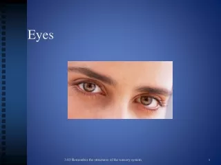
Vision • We rely on our vision more than any other special sense • Visual receptors are located in the eye
Functions of accessory structures • Protection • Lubrication • Secretion of tears
Accessory structures of the eye • Superficial Epithelium of the Eye- thin layers of skin around the eye and covering the eye itself • Eyelashes- robust hairs that help prevent foreign materials from reaching the eye • Eyelids – continuation of the skin that protect and lubricate the eye
eyelashes • Located along the inner margin of the eye lid • Tarsal Glands- also known as Meibomian, are modified sebaceous glands • Tarsal glands secrete lipid-rich products that keep the eye lids from sticking together
Eyelids • Eyelids open and close eye using muscles fibers • Orbicularis Oculi and Levator Palpebrae Superioris muscles are responsible for closing the eye and raising the upper lid
Epithelium of the eye • Conjunctiva- outer surface of the eye that a mucous membrane covered in stratified squamous epithelium • Palpebral Conjunctiva- inner surface of the eyelid • Ocular Conjunctiva- the anterior surface of the eye • Cornea- a transparent part of the outer fibrous layer
Lacrimal apparatus • Lacrimal Apparatus- produces, distributes, and removes tears • Consists of • Lacrimal Gland and associated ducts • Lacrimal Canaliculi • Lacrimal Sac • Nasolacrimal Duct
Lacrimal apparatus • Lacrimal Gland- tear gland • Lacrimal Canaliculi- small canals that lead to the lacrimal sac • Lacrimal Sac-holds the tears that the lacrimal gland produces • Nasolacrimal Duct- delivers tears to the nasal cavity on that side
The eye • Sophisticated visual instruments • Contains three distinct layers or tunics • Outer Fibrous Tunic • Middle Vascular Tunic • Inner Neural Tunic (retina)
Fibrous tunic • Outermost layer • Consists of sclera and cornea • Sclera- “white of the eye”; made of collagen and elastic fibers • Provides mechanical support and some physical protection • Serves as an attachment site for the eye muscles • Contains structures that assist in the focusing process
Vascular tunic • Also known as the Uvea • Contains blood vessels, lymphatic vessels, and the intrinsic muscles of the eye • Provides a route for blood vessels and lymphatics that supply tissues of the eye • Regulating the amount of light the eye receives
Vascular tunic • Secreting and reabsorbing the aqueous humor that circulates the eye • Controls the shape of the lens • Contains the iris • Visual receptors, or Photoreceptors, located in neural tunic
Iris • Iris- visible through the corneal surface, contains the blood vessels, pigment cells, and smooth muscle fibers • Pupillary muscles- muscles that contract and changes the diameter of the pupil • Pupil- central opening of the iris
Pupillary muscles • Pupillary Constrictor Muscles- when it contracts, the pupil decreases (more light) • Pupillary Dilator Muscles- contraction enlarges the pupil (less light)
Neural Tunic • Also known as the Retina • Retina helps process visual information • Contains two parts: pigmented part and neural part • Pigmented part absorbs light • Neural part is in control of processing • Also contains photoreceptors • Photoreceptors- cells that detect light
Organization of retina • Rods and cones • Rods- highly sensitive to light, don’t ‘see’ colors • Cones- ‘sees’ colors, provide sharper clearer images • Optic Nerve- transmits the visual images picked up from the rods and cones and delivers them to the brain
Rods and cones • Macula Lutea- has no rods • Fovea- contains highest cone concentration • Fovea is the site of the sharpest vision
Structure of the eye • The eye is hollow • Two cavities • Posterior cavity • Anterior cavity is filled with aqueous humor
Posterior cavity • Or Vitreous Chamber, contains the vitreous body • Vitreous Body- or Vitreous Humor, gelatinous substance that makes up most of the volume of the posterior cavity • Helps stabilize the shape of the eye
Anterior Cavity • Divided into two chambers • Anterior chamber • Posterior chambers • Chambers are filled with Aqueous Humor • Aqueous Humor- fluid that circulates within the anterior cavity, passing through the chambers of the pupil
Anterior chamber • Extends from the cornea to the iris
Posterior chamber • Extends between the iris and the lens
lens • Lies posterior to the cornea • Primary function is to focus the visual image on the photoreceptors • Focus happens by the change in shape of the lens • Lens fibers are in the interior of the lens
Lens fibers • Lost their nucleus and organelles • Slender and elongated • Filled with transparent proteins called crystallins • Crystallins- responsible for clarity and focusing power of the lens
transparency • Depends on precise combination of structural and biochemical characteristics • Lose of balance produces cataracts
refraction • The light that is collected by the photoreceptors in refracted, or bent when passing from one medium to another • Pencil in water • Refraction occurs when passing light through the cornea and then into the lens
refraction • Greatest amount of refraction occurs when light passes through the air into the corneal tissues • Tissues have a density similar to water • When you opne your eyes underwater you cant see as easily because the air-water refraction has been eliminated and replaced with water to water, thus light remains unbent and
Additional refraction • Light passes through the aqueous humor into the dense lens • This lens provides extra refraction that’s needed to focus the light rays from an object to a focal point • Focal Point- a specific point of intersection of the retina
Focal distance • Focal Distance- distance between the center of the lens and its focal point • Determined by two factors • Distance from object to the lens • Shape of the lens
Distance from the object to the lens • The closer an object is to the lens, the greater the focal distance
The shape of the lens • The rounder the lens the more refraction occurs, so a very round lens has a shorter focal distance than a flatter one
accommodation • Accommodation- focusing images on the retina by changing the shape of the lens to keep the focal length constant • To view nearby objects the lens becomes rounder • The lens flattens when we view a distant object • Lens are held in place by suspensory ligaments
accommodation • Greatest amount of refraction is needed for viewing objects up close • Inner limit of clear vision is called the near point of vision • Children can see things up close but as time goes on the lens becomes stiffer and less responsive • Aging effects the near point of vision
Astigmatism • If light doesn’t pass properly the image is distorted • Astigmatism- the degree of curvature in the cornea or lens varies from one axis to another • Image distortion may be so minimal people don’t even notice the condition
Image reversal • Light originates at a single point either near or far • However and object in view is a complex light source that is treated as a large number of individual points • These individual points creates a miniature image of the original but is upside down and backwards • The brains compensates for this image reversal and we don’t notice it
Visual activity • Visual activity- clarity of vision • Rated against a ‘normal’ standard (20/20, 20/15, etc.) • Considered legally blind when vision falls below 20/200, even with glasses or contact lenses
blindness • Terms implies a total absence of vision due to damage of the optic pathways • Common causes are • Diabetes mellitus • Cataracts • Glaucoma • Corneal scarring • Detachment of the retina • Hereditary factors
Scotomas • Abnormal blind spots that may appear in the field of vision • Permanent in a fixed position • Result from a compression of the optic nerve, damage to the photoreceptors, of damage to the visual pathway • Also Floaters, which a small spots that drift across the field of vision, generally temporary phenomena
Color vision • Objects appear to have color if they reflect or transmit photons from one portion of the visible spectrum and absorbs the rest • Photons stimulate rods and cones • Photons of all colors bounce off an object or rods themselves are stimulated, the object will appear white • If photons are absorbed by the object (none reach the retina), the object appears black
Cone types • Blue cones, green cones, and red cones • Each have a sensitivity to a different range of wavelengths • Stimulation to different combos of wavelength creates color vision • Color discrimination results from the integration of info from all three types of cones • EXAMPLE: Yellow is formed from a combo of highly stimulate green cones, less strongly stimulated red cones, and relatively unaffected blue cones