Download
1 / 63
630 likes | 818 Views
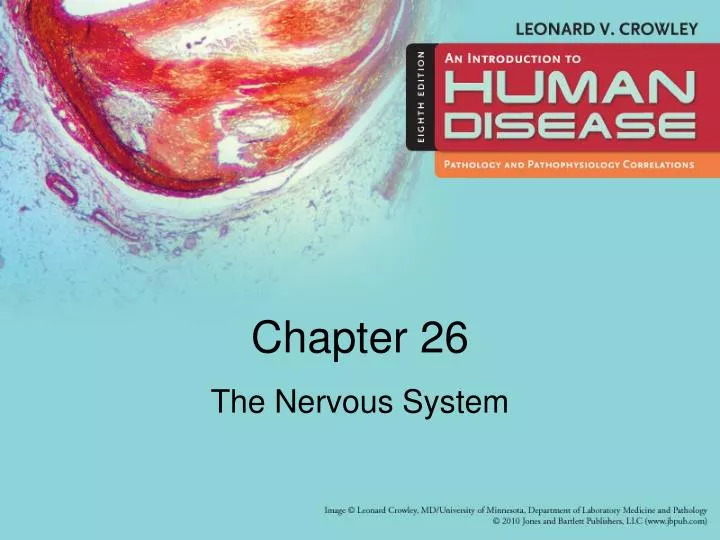
Chapter 26. The Nervous System. Learning Objectives (1 of 2). Describe normal structure and function of the brain, meninges, CSF in relation to neurologic disease Define muscle tone, voluntary motor activity and relate to two forms of muscle paralysis

E N D
Chapter 26 The Nervous System
Learning Objectives (1 of 2) • Describe normal structure and function of the brain, meninges, CSF in relation to neurologic disease • Define muscle tone, voluntary motor activity and relate to two forms of muscle paralysis • Explain pathogenesis, prenatal diagnosis, clinical manifestations of closure defects of the CNS • Describe pathogenesis, clinical manifestations of hydrocephalus and relate to treatment measures • Describe causes, manifestations, treatment of transient ischemic attack, TIA
Learning Objectives (2 of 2) • Differentiate types of stroke as to pathogenesis, prognosis, and treatment • Describe pathogenesis, manifestations, treatment of congenital cerebral aneurysms • Explain pathogenesis, origin, clinical manifestations, and treatment of CNS tumors • Explain pathogenesis, clinical manifestations, and treatment of Parkinson’s disease, meningitis, multiple sclerosis, and Guillian Barre syndrome
Nervous System • Central nervous system, CNS • Brain • Spinal cord • Meninges: surrounding membranes • Neurons (nerve cells) and neuroglia (supporting cells) • Sensory or afferent nerve: transmits impulses to the nervous system • Motor or efferent nerve: transmits impulses from brain or spinal cord to muscle • Transmission of a nerve impulse via neurotransmitters • Acetylcholine, norepinephrine, dopamine
Meninges • Dura: firm, outer covering • Arachnoid: middle • Subarachnoid space: space between arachnoid and pia contains • CSF (cerebrospinal fluid) • Strands of arachnoid connective tissue • Pia: thin, inner membrane • Adheres to brain and spinal cord
Brain • Cerebrum • Cerebellum • Brain stem • Brain: four cavities called ventricles • Tissues of brain and spinal cord • Nerve cells = neurons • Supporting cells = neuroglia • Arterial blood supply • Large vessels enter base of skull • Vessels join to form arterial circle at base of brain • Venous blood • From brain into large venous sinuses in dura • Sinuses eventually drain into jugular veins
Development of Nervous System • Neural plate becomes neural tube • Forebrain forms cerebral hemispheres and diencephalon • Midbrain and hindbrain form remainder of adult brain • Mesoderm surrounding neural tube forms cranial cavity, vertebral bodies, and surrounding structures
Voluntary Motor Activity • Controlled by nerve impulses originating in motor neurons of the cerebral cortex (cortical neurons) • Muscle tone caused by reflex arcs • Pyramidal system controls voluntary motor functions • Extrapyramidal system regulates muscle groups concerned with automatic functions such as walking
Muscle Paralysis • Flaccid paralysis • Destruction of motor neurons by disease • Interruption of reflex arc responsible for muscle tone • Muscle deprived of innervation • Low muscle tone • Peripheral nerve destruction • Spastic paralysis • Reflex arc not disturbed • Injury to cortical neurons stops voluntary control • Muscle retains innervation • Increased muscle tone
Cerebral Injury • Large blood vessels over surface of brain may be torn by force of injury • Epidural hemorrhage • Subdural hemorrhage • Subarachnoid hemorrhage
Mechanism of injury to frontal and temporal poles of brain caused by blow to back of head.
Skull x-ray illustrating large skull fracture (arrows) associated with extensive injury to underlying brain.
Neural Tube Defects • Anencephaly • Failure of normal development of brain and cranial cavity • Multifactorial inheritance • Spina bifida • Diagnosis: amniocentesis and alpha-fetoprotein levels • Alpha-fetoprotein leaks from fetal blood into amnionic fluid through open neural tube defect; high levels found in amnionic fluid • Occult • Meningocele • Meningomyelocele
Characteristic appearance of anencephalic infant. Lateral view.
Neural Tube DefectsA. Thoracic meningomyelocele covered by thin membraneB. Large meningomyelocele associated with neurologic deficit
Hydrocephalus • Congenital hydrocephalus • From congenital obstruction of aqueduct or absence of openings in roof of 4th ventricle • Head enlarges as ventricles dilate because cranial structures have not fused • Acquired hydrocephalus • Obstruction of CSF by tumor or adhesions blocking opening in 4th ventricle • Ventricles dilate but head does not enlarge because cranial structures are fused
Coronal section of brain revealing marked dilation of ventricles in patient with congenital hydrocephalus.
Stroke: Cerebrovascular Accident (1 of 5) • Any injury to brain tissue from disturbance of blood supply to brain • Types of stroke • Cerebral thrombosis: most common; thrombosis of cerebral artery narrowed by arteriosclerosis • Cerebral embolus: occurs less frequently; blockage of cerebral artery by fragment of blood clot from an arteriosclerotic plaque or from heart • Cerebral hemorrhage: most serious type of stroke; usually from rupture of a cerebral artery in person with hypertension
Stroke: Cerebrovascular Accident (2 of 5) • Predisposing Factors • 1. Mural thrombus formed on wall of left ventricle adjacent to a healing myocardial infarction • 2. Thrombus formed on rough surface of diseased mitral or aortic valve • 3. Small thrombus in left atrium of person with atrial fibrillation
Stroke: Cerebrovascular Accident (3 of 5) • Ischemic infarct: no blood leaks into brain • Hemorrhagic infarct: blood leaks into damaged brain tissue • Arteriosclerosis of extracranial arteries • Sclerosis of a major artery from aorta that supply brain • Common affected site: carotid artery in neck; arteriosclerotic plaque may narrow lumen and reduce cerebral blood flow
Stroke: Cerebrovascular Accident (4 of 5) • Diagnosis • Cerebral angiogram • Carotid endarterectomy • Less invasive methods: similar to balloon angioplasty and stent insertion procedures used to treat coronary artery plaques
Stroke: Cerebrovascular Accident (5 of 5) • CT scan: can distinguish a cerebral infarct from cerebral hemorrhage • Magnetic resonance imaging (MRI): provides similar information and is equally effective
An angiogram revealing narrowing of the carotid artery in the neck (arrows).
Infarct, right cerebral hemisphere from thrombosis of middle cerebral artery
Effects of atherosclerosis of carotid arteryA. Narrowing of lumenB. Thrombus formationC. Thrombus dislodged & forms emboliD. Complete occlusion of artery by thrombus
Carotid endarterectomyA. Resected atherosclerotic plaque follows contour of common carotid artery and branchesB. Opened endarterectomy specimen
Coronal section of brain illustrating large cerebral hemorrhage that has compressed and displaced the cerebral ventricles.
A computed tomographic (CT) scan of a patient with cerebral hemorrhage (arrow), which appears white because blood is denser than brain tissue.
Transient Ischemic Attack, TIA • Brief episodes of neurologic disfunction • From embolization of material from plaque in carotid artery • One-third of patients eventually suffer major stroke • Treatment: endarterectomy or medical therapy
Cerebral Aneurysm • Congenital aneurysm of circle of Willis • Congenital weakness in arterial wall allows lining to protrude • Weakness is congenital but aneurysm develops in adult life • Rupture causes subarachnoid hemorrhage • Hypertension predisposes • Treatment: aneurysm occluded surgically • Arteriosclerotic aneurysm • Cerebral artery dilates and compresses adjacent tissue • Rupture uncommon
Dissection of vessels from the brain of a person with large congenital cerebral aneurysm.
A large ateriosclerotic aneurysm (arrows) that compressed and distorted the brain stem.
Undersurface of brain, illustrating subarachnoid hemorrhage secondary to ruptured cerebral aneurysm.
Infections • Three types • Bacterial • Fungal • Viral • Meningitis: infection affecting meninges • Encephalitis: infection of brain tissue • Meningoencephalitis: affects both meninges and brain tissue
Meningitis from Bacteria and Fungi • Meningococcus (Neisseria meningiditis) • Pneumococcus (Streptococcus pneumoniae) • Hemophilus influenzae
Bacterial meningitis, illustrating purulent exudate in the meninges
Viral Infections That Affect the CNS (1 of 2) • Measles, mumps, herpes simplex virus, intestinal and respiratory viruses, cytomegalovirus, poliomyelitis virus, and arborviruses • Manifestations • Systemic symptoms • Aseptic meningitis: caused by a virus • Suppurative meningitis: pus-producing; caused by bacteria • Encephalitis: brain tissue involvement • Spinal fluid abnormalities
Viral Infections That Affect the CNS (2 of 2) • Arboviruses: responsible for cases of meningitis and encephalitis • Viruses infect birds, animals, humans; transmitted by mosquitoes • Types of encephalitis • Western equine encephalitis • Eastern equine encephalitis • St. Louis encephalitis • California encephalitis • West Nile virus: “foreign” virus from Africa, first case identified in 1999 in New York City area
Creutzfeldt-Jakob Disease • Caused by small protein particle produced as a result of gene mutation • Normal form of protein: “good prion” designated as PrPc • Abnormal form: “bad prion” designated as PrPsc • Mad cow disease • Prion disease affecting cows • Cows become infected from animal feed mixed with protein-rich tissue from sheep infected with scrapie • Eating infected beef causes variant Creutzfeldt-Jakob disease in humans
A photomicrograph of cerebral tissue from patient with Creutzfeldt-Jakob disease.
Alzheimer Disease • Characteristics • Progressive mental deterioration • Emotional disturbances • Anatomic and biochemical features • Neurofibrillary tangles: thickening of neurofilaments • Neurotic plaques: clusters of thick, broken neurofilaments • Biochemical abnormalities and brain enzyme deficiencies: acetylcholine and acetylcholine synthesizing enzyme • No specific treatment; some drugs may temporarily improve cerebral function
Alzheimer DiseaseThickened neurofilaments forming neurofibrillary tangles
Multiple Sclerosis • Probably an autoimmune disease in generally predisposed individual • Random foci of demyelination followed by glial scarring • Neurologic symptoms depend on location of plaques • Probably initiated by a viral infection in a genetically predisposed person • Manifestations • Activated T lymphocytes, monocytes target myelin proteins, destroy myelin • Treatment • MRI demonstrates plaques in CNS
Coronal section of brain illustrating areas of gilial scarring (arrows) adjacent to ventricle in multiple sclerosis.