Download
1 / 23
260 likes | 621 Views
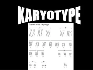
Chicken karyotype analysis/ SCEs. Ciaran Morrison. Diagrammatic representation of the chicken karyotype. The DT40 karyotype. Modal karyotype is: 2 Gga-1, 3 Gga-2, 2 Gga-3, 2 Gga-4, 2 Gga-5, 1 Gga-Z. From: Sonoda et al. (1998) EMBO J. 17 : 598-608.

E N D
Chicken karyotype analysis/ SCEs Ciaran Morrison
The DT40 karyotype Modal karyotype is: 2 Gga-1, 3 Gga-2, 2 Gga-3, 2 Gga-4, 2 Gga-5, 1 Gga-Z. From: Sonoda et al. (1998) EMBO J. 17: 598-608
Variation/ mosaicism in the DT40 karyotype Variations observed in karyotypes during extended culture periods, within the same cultures. Particular variations included: 2 Gga-2, 1 Gga-4; 1 Gga-3; 1 Gga-1 Chang & Delany (2004) Chromosome Res. 12: 299-307 From: Sonoda et al. (1998) EMBO J. 17: 598-608
Metaphase spreads-basics • Block cells in metaphase (colcemid for up to 4h; longer results in v. high condensation) • Hypotonically swell cells. • Fix cells and drop on to slides. • Dry and stain with Giemsa (or DAPI)
5 2 3 1 5 2 3 4 Z 2 1 4 Anna Stephan
Chromosome aberrations From: Sonoda et al. (1998) EMBO J. 17: 598-608
Chromosome aberrations Analysis allows the observation of DNA damage in chromosomes. Analysis may indicate specific repair deficiencies or abnormalities (e.g., abnormalities in both sisters is often from post-replicative repair, i.e. HR)
‘A Spotter’s Guide’ Ctg: Chromatid-type gap Ctb: Chromatid-type break Csg: Chromosome gap Csb: Chromosome break Cte: Chromatid exchange
Scoring suggestion Record data for each macrochromosome in a spread. Sum for population (100 metaphases)
Telomere analysis in DT40s Chickens have high levels of telomere repeat sequence, notably in the microchromosomes. DT40s have shorter-than-normal telomeres, although the interstitial sequences are retained. From: O’Hare & Delany (2009) Chromosome Res. 17: 947-64.
Recent information on centromeres in DT40s • Shang WH, Hori T, Toyoda A, Kato J, Popendorf K, Sakakibara Y, Fujiyama A, Fukagawa T. • Genome Res. 2010 Jun 9. [Epub ahead of print] • The chicken genome has a hybrid centromere model, involving either long arrays of tandem repeats on some chromosomes or relative short spans of non-tandem-repeat sequences on other chromosomes.
Sister chromatid exchanges • Equal exchanges between sister chromatids (post-replicative). • Reflect homologous recombinational repair activities in cells. • Frequently used in mutagenesis/ toxicity assays as readouts for environmental stressors. • Greatly elevated in certain human diseases (Bloom’s syndrome).
Sister chromatid exchanges Sonoda, E. et al. 1999. Mol. Cell. Biol. 19(7):5166-5169
SCE visualisation + BrdU 1 cell cycle Equally-labelled sisters
SCE visualisation + BrdU 2nd cell cycle Differentially-labelled sisters
SCE visualisation + BrdU 2nd cell cycle Differentially-labelled sisters Giemsa staining reveals differential labelling
SCE visualisation + BrdU 2nd cell cycle Differentially-labelled sisters Giemsa staining reveals differential labelling and exchanges
SCEs: observation • Requires labelling for 2 cell cycles; optimise! • Visualisation of differential labelling may include Hoechst staining (to magnify differences between the labelled/ unlabelled sisters) Fluorescence plus Giemsa (‘FPG’) Simpson, LJ and Sale, JE (2006) ‘Sister chromatid exchange assay’, in Buerstedde and Takeda (eds.) Reviews and Protocols in DT40 Research 399-403.