Download
1 / 35

E N D
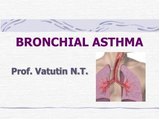
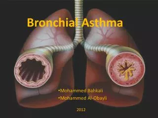
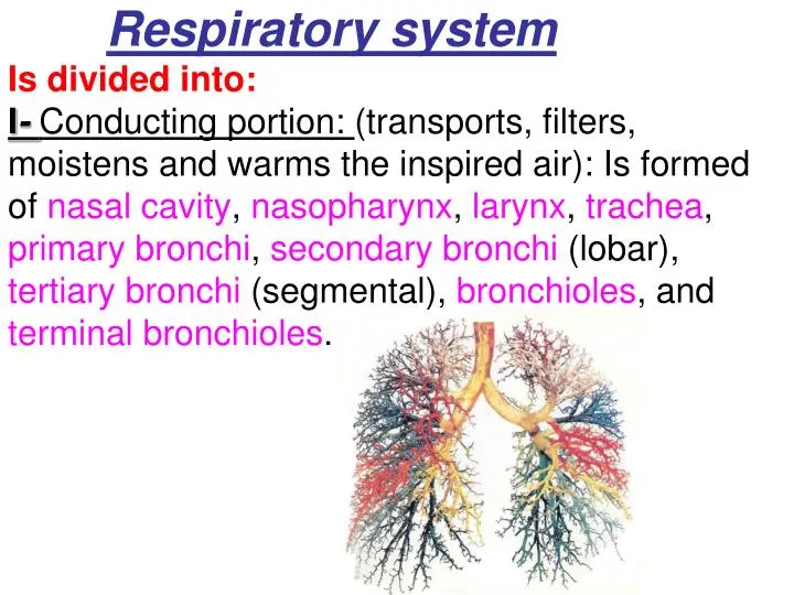
Respiratory systemIs divided into:I- Conducting portion: (transports, filters, moistens and warms the inspired air): Is formed of nasalcavity, nasopharynx, larynx, trachea, primary bronchi, secondary bronchi (lobar), tertiary bronchi (segmental), bronchioles, and terminal bronchioles.
II- Respiratory portion: ( has alveoli- the sites of gas exchange): Is formed of respiratorybronchioles, alveolar ducts, alveolar sacs and alveoli.
Bronchial Tree • Begins at thebifurcation of the trachea • Is composed of: • Extrapulmonary Part: • Primary (main) bronchi. • Intrapulmonary Part: that includes: • Secondary (lobar) bronchi • Tertiary (segmental) bronchi • Bronchioles • Terminal bronchioles • Respiratory bronchioles. • Alveolar Ducts and sacs • Alveoli
Trachea • It is formed of: • 1.Mucosa, composed of: * Lining epith. • a-Respiratory epith. • b-Basal cells. • c-Brush cells. • d-Serous cells. • e-DNES (defuse neuro-endocrine cells). • Lamina propria: fibroelasticC.T.contains lymphoid elements, mucous, serous glands and elastic lamina.
2.Submucosa: is composed of denseirregular fibero-elastic C.T. containing mucous and seromucousglands. It contains lymphoid elements and bl. Vessels.3.Adventitia:is composed of fibero-elastic C.T.and C-rings of hyaline cartilage.
General Characters of Bronchial Tree • Progressive decrease in: • Size (diameter) • Amount of cartilage. • Number of glands and goblet cells • The height of epithelial cells. • Progressive increase in: • Amount of smooth muscle • Amount of elastic tissue
Primary bronchi are identical in structure to trachea (but smaller).Intrapulmonary bronchi have irregular plates of hyaline cartilage (in lieu of C-ring), which completely surrounds the lumen of intrapulmonary bronchi.
Bronchioles. • Are less than 1mm in diameter. • Have no cartilage. • Have Clara cells. • Large bronchioles are lined with simple columnar ciliated epithelium with few goblet cells. • Small bronchioles are lined with simple cuboidal ciliated epith.without goblet cells. • Lamina propria has NO glands. • Are surrounded by helically oriented smooth muscle layers that surrounded by elastic fibers.
Clara cells • Are columnar cells with dome shaped apices. • Have microvilli. • Have abundant rER. • Are secretory cells, (glycoprotein and surfactant-like material) • Have abundant sER. • Can degrade toxins in the inhaled air. • Can divide to regenerate the bronchiolar epith.
Terminal Bronchioles. • Are the smallest and most distal region of the conducting portion (less than o.5mm). • Are lined by Clara cells and partially ciliated cuboidal cells. • Lamina propria is formed of fibro-elastic C.T. • Have one or two layers of smooth muscle. • Elastic fibers radiate from the adventitia. • They give rise to respiratory bronchioles.
Respiratory bronchioles • Have the same structure as terminal bronchioles but, • They have alveoli through them gas exchange takes place. • They terminate in an alveolar ducts.
Alveolar ducts • Do not have walls of their own, but are lined with extremely attenuated squamous alveolar cells. • They end with two or more small clusters of alveoli (alveolar sac). • The opening of each alveolus to the alveolar duct is controlled by a single smooth muscle cell, embedded in type III collagen. • Fine elastic fibers ramify from the alveolar ducts and sacs.
Alveolus • Is a small outpouching of respiratory bronchiole, alveolar ducts and alveolar sacs. • Is the structural and functional unit of respiratory system. • Contiguous alveoli communicate through an alveolar pore of Kohn (equilibrate air pressure). • Interalveolar septum is occupied by extensive continuous blood capillaries and C.T. rich in elastic and type III collagen. • Opening of alveoli associated with alveolar sacs are devoid of smooth muscle cells, instead there are elastic and reticular fibers.
Lining of the alveoli • A-Type I pneumocytes : *Line about 95% of alveolar surface. *Highly attenuated squamous cells. *Have thin cytoplasm that contain few mitochondria, few rER and a modest Golgi apparatus. *Have occluding junctions. *have well developed basal lamina.
B-Type II pneumocytes*occupy 5% of the alveolar surface.*Are cuboidal cells.*Form occluding junctions with type I alveolar cells.*Occupy most of the alveolar septa.*Have basal lamina.*Have apical microvilli.*Have central nuclei.*Have abundant rER, well developed Golgi apparatus and mitochondria.*Have membrane-bound lamellar bodies.*Secrete pulmonary surfactant (phospholipids and proteins) and undergo mitosis.
The surfactant • Is secreted by exocytosis from type II alveolar cells into the lumen of the alveolus. • Decrease surface tension of the alveoli thus prevent their collapse.
EM of Blood-Gas Barrier *Pneumocytes type I and surfactant. *Fused basal lamina of type I pneumocytes and endothelial cells of the capillary. *Endothelial cells of the continuous capillaries.
Interalveolar Septum • Lies between the alveoli • Contains extensive continuous blood capillaries, Dust cells, macrophages and CT rich in elastic and type III collagen • Contiguous alveoli communicate through an alveolar pore of Kohn to equilibrate air pressure.
Alveolar macrophages (dust cells) • Are monocytes in the pulmonary interstitium. • Migrate between type I alveolar cells, and enter the alveoli. • Phagocytose dust and bacteria. • Migrate to bronchi to the pharynx or into lymph vessels.
PleuraIs formed of two layers:Parietal and visceral.It is formed ofsimple squamous mesothelium. The two layers are separated by serous of fluid. The visceral layer has sub-epithelium loose C.T that extends in lung tissue