Download
1 / 16
160 likes | 295 Views
Cortico -spinal tract integrity measured using magnetic resonance imaging and transcranial magnetic stimulation in n euromyelitis optica and multiple sclerosis.

E N D
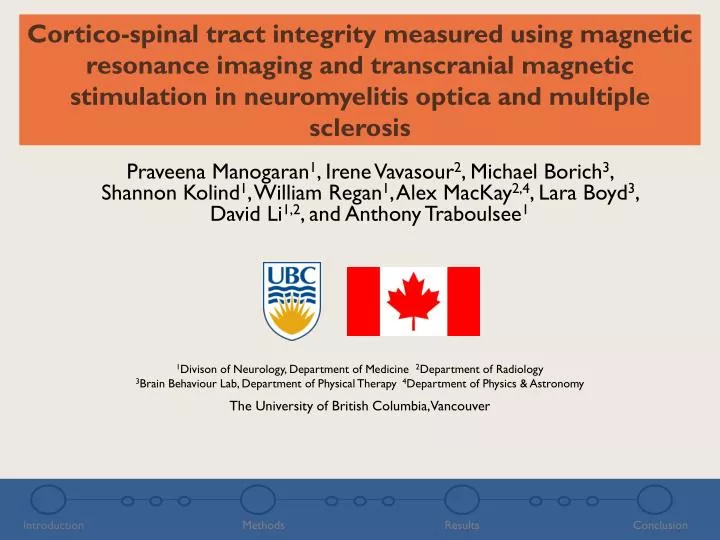
Cortico-spinal tract integrity measured using magnetic resonance imaging and transcranial magnetic stimulation in neuromyelitis optica and multiple sclerosis Praveena Manogaran1, Irene Vavasour2, Michael Borich3, Shannon Kolind1, William Regan1, Alex MacKay2,4, Lara Boyd3, David Li1,2, and Anthony Traboulsee1 1Divison of Neurology, Department of Medicine 2Department of Radiology 3Brain Behaviour Lab, Department of Physical Therapy 4Department of Physics & Astronomy The University of British Columbia, Vancouver Introduction Methods Results Conclusion
Multiple Sclerosis (MS) Central: Fatigue Cognitive Impairment Depression Visual: Optic neuritis Diplopia Axon Myelin Musculoskeletal: Weakness Spasms Ataxia Sensation: Pain Bowel: Incontinence Diarrhea Damaged Myelin Urinary: Incontinence Frequency Retention T2-weighted - 4 hyperintense lesions http://www.rethinkmsrelapses.com/pages/understand_ms_relapses/what_causes_an_ms_relapse Barkhof F et al. Brain 1997; 11: 2059-2069 Introduction Methods Results Conclusion
Neuromyelitis Optica (NMO) • Characterized by damage to astrocytes • Usually presents with severe axonal degeneration • Shows similar clinical and radiological features to MS but treatment and prognosis differ significantly Myelitis Optic Neuritis http://www.unitedspinal.org/msscene/2008/02/04/neuromyelitis-optica/ Introduction Methods Results Conclusion
Objective 1. Characterize differences incortical excitabilityand myelin status of descending motor output pathways 2. Evaluate the relationships between these measures in individuals with MS and NMO compared to healthy controls Introduction Methods Results Conclusion
Methods Introduction Methods Results Conclusion
Study Population • 30 subjects age and gender matched • MRI protocol performed on Philips 3.0T Achieva system Introduction Methods Results Conclusion
T2 Relaxation and Myelin Water Fraction 40 ms 500 ms T2 relaxation of MRI signal Amplitude 0 10 20 30 40 50 60 70 80 Signal Amplitude Fit T2 data with NNLS to obtain Time (ms) 10 100 1000 T2 Time (ms) Images courtesy of Sandra Meyers, UBC MRI Research Group Introduction Methods Results Conclusion
Transcranial Magnetic Stimulation (TMS) Motor Evoked Potential (MEP) Image courtesy of the Brain Behaviour Lab • Figure-of-eight coil attached to a Magstim 200 stimulator targeting motor cortex TMSis a non-invasive, safe, and quick measure of cortical-spinal excitability MEPs recorded with surface electromyography of extensor carpi radialis bilaterally Introduction Methods Results Conclusion
Transcranial Magnetic Stimulation (TMS) Strafella AP & Paus T.J Neurophysiol2001; 85(6): 2624-2629 MEP Test Stimulus Test Response 2 msinterstimulusinterval = Intracortical Inhibition (ICI) 12 msinterstimulusinterval = Intracortical Facilitation (ICF) PP ratio = conditioned MEP/unconditioned mean MEP Paired pulse TMS evaluates ICI and ICF pathways Introduction Methods Results Conclusion
RESULTS Introduction Methods Results Conclusion
Myelin water fraction is decreased in neuromyelitis optica compared to multiple sclerosis and healthy controls ** p < 0.01*** p < 0.001 Introduction Methods Results Conclusion
Lower intracortical inhibition in multiple sclerosis compared to neuromyelitis optica and healthy controls ** p < 0.01 Introduction Methods Results Conclusion
No significant correlations were found between myelin water fraction and intracortical excitability measures Introduction Methods Results Conclusion
Conclusions • Structural changes found in the descending motor output pathway white matter of NMO patients • Cortical excitability changes were observed in MS patients that were specific to intracortical inhibitory pathways • Intracortical inhibition may not be directly linked to myelination Introduction Methods Results Conclusion
Conclusions These neurophysiological and neuroanatomical changes may offer a novel biomarker to distinguish between individuals with MS and NMO Introduction Methods Results Conclusion
Acknowledgements Brain Behaviour Lab Michael Borich MarjanZakeri Lara Boyd Tamara Koren Andrej Satara MS/MRI Research Group William Regan Annie Kuan Rachel Kim Study subjects MRI Research Group Anthony Traboulsee Shannon Kolind Irene Vavasour Sandra Meyers Alex MacKay Eric Zhao Nolan Shelley David Li Alex Rauscher UBC’s MRI Technologists