Download
1 / 18
180 likes | 871 Views
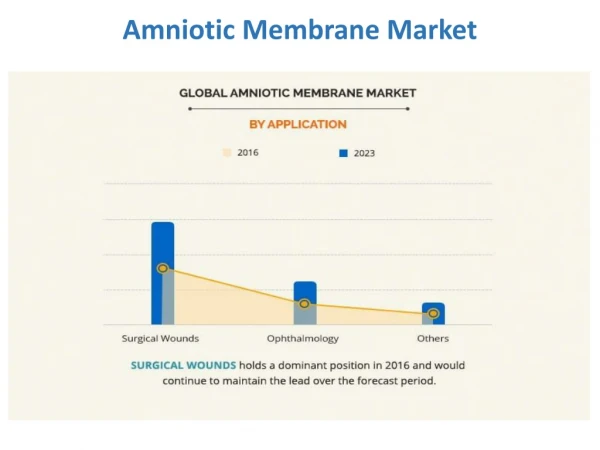
Purpose . Conjunctivochalasis, defined as a redundant conjunctiva typically located between the globe and the lower eyelid, is not uncommon.The objective of this article is to report surgical outcomes of conjunctival resection and amniotic membrane transplantation on conjunctivochalasis. . Methods.

E N D
1. Surgical Outcomes of Conjunctival Resection and Amniotic Membrane Transplantation onConjunctivochalasis Ji-Woong Lee, MD, Byung-Ryong Choi, MD, Hong-Kyun Kim, MD,1 Hee-Tae Cho, MD.2
1Department of Ophthalmology, Kyungpook National University College of Medicine,
2Cho Hee Tae eye clinic, Daegu, Korea
3. Methods The authors studied 4 patients (8 eyes) of conjunctivochalasis from April 2004 to July 2005(Table 1).
There was no lid abnormality such as lid laxity, trichiasis, entropion, ectropion in all patients.
We did not observe any outflow obstruction by lacrimal irrigation test.
All patients had redundant conjunctiva typically located between the globe and the lower eyelid and had delayed tear clearance measured by fluorescein clearance test.
4. They were given artificial tear substitutes and corticosteroid eyedrops.
But, they were refractory�to drug therapy.
Operation was performed on both eyes under the general anesthesia.
We marked with Gentian Violet after grasping redundant conjunctiva with forcep and resected it in crescent shape along the plica semilunaris.
We cover the defect with amniotic membrane using 8-0 vicryl.
5. Postoperatively, all patients received 0.1% ofloxacin eyedrops and 0.1% fluorometholone eyedrops four times daily for 2 weeks.
The sutures were removed 2 weeks after the operation and we checked the resolution of the symptoms and development of complications in all patients.
6. Results The subject�s ages ranged from 56 to 74 years. They complained of epiphora and foreign body sensation and were found to have redundancy of conjunctival tissue. They showed delayed tear clearance measured by fluorescein clearance test (Table 1).
7. Representative case shows redundant conjunctiva which covers the inferior limbus and inferior part of the cornea and it disturbs the formation of an adequate tear meniscus preoperatively, but the conjunctiva near the lower eyelid is not redundant postoperatively (Fig. 1).
8. There show the redundant conjunctiva, conjunctival chemosis and conjunctival cyst in other patients preoperatively, but the conjunctiva is not redundant postoperatively (Fig.2).
There were improvement of symptoms such as epiphora, foreign body sensation and loss of redundant conjunctiva after surgical treatment.
After removing the suture, we observed no complication and cosmetic problems in all patients.
12. Discussion Possible mechanisms to form conjunctivochalasis are mast cell activation which destruct conjunctival architecture, senile change such as degeneration of elastic fibers, and degrading enzymes derived from tears accumulated in the inferior fornix and the tear meniscus.
13. Conjunctivochalasis can cause a spectrum of symptoms, ranging from aggravation of a dry eye at the mild stage, to disturbance of tear outflow at the moderate stage by interference of the inferior tear meniscus and occlusion of the inferior punctum, and exposure problems at the severe stage such as nocturnal lagophthalmos and dellen.
14. No treatment is needed for asymptomatic conjunctivochalasis.
Patients with severe conjunctivochalasis, who have ocular irritation, pain, ulceration,or subconjunctival hemorrhage, may be given tear substitutes, lubricants, and corticosteroid or antihistamine drops and be advised to patch before sleep to reduce nocturnal exposure.
If these measures are unsuccessful, surgical treatment becomes necessary.
15. The term conjunctivochalasis, taken from the Greek term meaning �relaxation of conjunctiva,� was first coined by Hughes in 1942.
The most commonly described method is a simple crescent excision of inferior bulbar conjunctiva 5 mm away from the limbus, followed by closure with absorbable sutures.
A modified technique, proposed to avoid visible scarring or retraction of the inferior conjunctival fornix includes peritomy made close to the limbus, after which two radial relaxing incisions to excise the redundant conjunctiva are made.
16. With either method, overzealous excision of conjunctiva may result in wound healing�induced complications, such as visible scarring, cicatricial entropion of the lower lid, retraction of the lower fornix, restricted motility, and corneal problems.
To obtain adequate excision yet avoid such complications, amniotic membrane transplantation has been introduced as a new surgical technique to reconstruct the inferior bulbar conjunctiva after excision (Fig. 3A,B).
Amniotic membranes stimulate differenciation and proliferation of conjunctival cell, suppress scar formation and inflammation.
18. Conclusions With minimum follow-up period of 2 months, symptoms resolved in most patients after removing the suture.
With amniotic membrane transplantation, we could avoid the complications that may result from overzealous resection of the conjunctival tissue, even though amnioitc membrane transplanation require more time than other surgical methods.
In conclusion, we successfully treated conjunctivochalasis with simple conjunctival resection and amniotic membrane transplantation and further study is needed.
And we emphasize the importance of careful examination of conjunctivochalasis during ocular examination of patients complaining tearing.