Download
1 / 24
240 likes | 551 Views
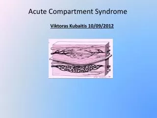
Compartment syndrome is a limb and life threatening condition that occurs when perfusion pressure falls below tissue pressure in a closed anatomical compartment .If left untreated -tissue necrosis and sequeleUltimately death It is found wherever a compartment is present.. Intro. Simple cause: T

E N D
1. By Suvarna Maharaj Compartment Syndrome- an overview
2. Compartment syndrome is a limb and life threatening condition that occurs when perfusion pressure falls below tissue pressure in a closed anatomical compartment .
If left untreated -tissue necrosis and sequele
Ultimately death
It is found wherever a compartment is present. Intro
3. Simple cause: THE PRESSURE IS TOO HIGH.
Either �decreased compartment size or increased fluid content.
Increased fluid content-
intensive muscle use
burns
intra-arterial injection
infiltrated infusion
haemorrhage
envenomation Causes
4. Decreased compartment pressure
Burns
Casts
Military aftershock trousers
Causes
5. This follows the path of ischemic injury. When fluid is introduced into a fixed volume or when volume decreases, pressure rises.
In the case of CS, compartments have a relatively fixed volume. An introduction of excess fluid or extraneous constriction increases pressure and decreases tissue perfusion until no O2 is available for cellular metabolism. Pathophysiology
6. Elevated perfusion pressure is the physiological response to rising intracompartmental pressure (IP). When IP rises, autoregulatory mechanisms are overwhelmed and a cascade of injury develops.
Tissue perfusion pressure is measured by subtracting the interstitial fluid pressure from the capillary perfusion pressure. When this pressure falls below a critical level, injury results. Pathophysiology cont.
7. When intracompartmentalpresssure rises, venous pressure rises. When venous pressure exceeds CPP, capillaries collapse. Generally, an intracompartmental pressure greater than 30mmHg requires intervention.
At this point, blood flow stops, resulting in decreased O2 delivery. Hypoxic injury causes cells to release vasoactive substances which increases endothelial permeability. Pathophysiology cont.
8. Capillaries allow continued fluid loss which increases tissue pressures and advances injury.
Nerve conduction slows,tissue ph falls due to anaerobic metabolism,surrounding tissue suffers further damage, and muscle tissue suffers necrosis releasing myoglobin.
The end is loss of the extremity and possibly, the loss of life. Pathophysiology cont.
9. Suspect CS whenever significant pain occurs in an extremity
Mechanism of injury- long bone fracture, high energy trauma, penetrating injuries, crush injuries
Remember to ask about anticoagulation-increases risk of CS Clinical- History
10. 5 P�s parasthesia, pallor,pulselessness, pain, poikilothermia are not diagnostic of CS. Except for pain and parasthesia , the other traditional signs are not reliable.
Severe pain at rest or with any movement especially passive stretching of the muscles should raise suspicion Signs
11. FOOT
-Classic signs What are they?
expected with foot fractures and injury so tense tissue bulging maybe the most reliable sign.
-associated with CS of deep posterior compartment of leg. Less common sites of CS
12. Symptoms from compression causes pain, loss of sensation and decreased hand function due to pressure on blood vessels and the median nerve within the wrist compartment . CS of the hand
13. The large gluteal muscle mass is confined in fascia hence area prone to CS. How?
Signs include pain especially on passive flexion at the hip and tense swelling of the buttock. Late signs include foot drop with a loss of sensation along distribution of sciatic nerve and no active movements of the ankle.
CS of the gluteal region
14. LAB STUDIES
Often normal and not helpful in diagnosing or excluding CS
Definitive diagnosis is compartment pressure measurement using a tonometer if available.
Remember PITFALLS Workup
15. Measurement Methods Simple needle
Wick Catheter
Slit catheter
Side Port catheter
Transducer �Tipped Catheter
16. Technique STRYKER TECHNIQUE
MERCURY MANOMETER
17. Technique
18. Go to www.emprocedures.com/compartment Demonstration
19. Stabilize the patient
Ischemic injury is basis for CS. Additional O2 should be given.
IV hydration is essential. Hypovolemia worsens ischemia.
Do not elevate the affected limb-decreases arterial pressure
Fasciotomy is definitive treatment so early referral is warranted. ED care
20. Two Incision Technique
Used to adequately decompress all four compartments
Medial Incision made longitudinally just posterior to tibia
Lateral incision made posterior to fibula from level of head to lat malleolus
Closure
Post-op Fasciotomies
21. Permanent nerve damage
Infection
Loss of limb
Death
Cosmetic deformity from fasciotomy Complications
22. Emedicine Compartment Syndrome by Richard Paula MD Director of Research, Assistant Professor of Emergency Medicine,University of South Florida
Mutimedia Procedure Manual- Compartment pressure Measurement
Gluteal Compartment Syndrome following Joint Arthroplasty Under Epidural Anaesthesia,Journal of Orthopaedics Surgery References
23. April 2007 By Kumar V Saeed, A Panagopoulos, PJ Parker
Wheeless� Textbook of Orthopaedics- Compartment syndrome of the Foot.
Acute Compartment Syndrome Update on Diagnosis and treatment by TE Whitesides and MM Heckman Academy of Orthopaedic Surgery July 1996
References
24. The end Thank you