Download
1 / 108
1.09k likes | 1.18k Views
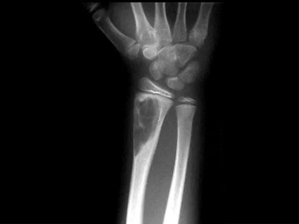
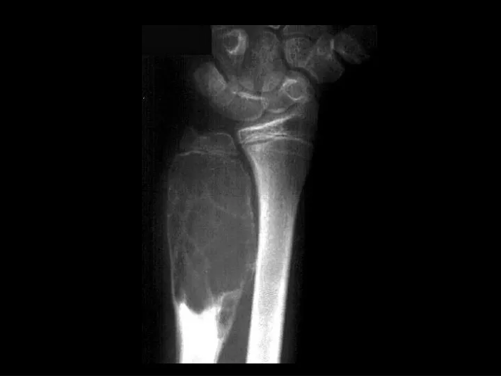
Aneurysmal Bone Cyst. Findings: expansile lytic lesion of the distal ulnar metaphysis well-defined margins internal septations No periostitis A result of trauma or tumor induced process ddx: Giant cell tumor Chondroblastoma Osteoblastoma Fibrous Dysplasia lytic met.

E N D
Aneurysmal Bone Cyst • Findings: • expansile lytic lesion of the distal ulnar metaphysis • well-defined margins • internal septations • No periostitis • A result of trauma or tumor induced process • ddx: • Giant cell tumor • Chondroblastoma • Osteoblastoma • Fibrous Dysplasia • lytic met
Osteopetrosis(Marble bones, Albers- Schoenberg) • Findings: • generalized increased bone density • rare bone dysplasia • Trabecular --> compact • Anemia w/extramedullary hematopoiesis • ddx: (children) • pyknodysostosis • renal osteodystrophy • vit A, D • Lead • Fluorosis
Paget’s Disease • Findings: • Enlargement of the right femoral head with course and thickend trabecula • Asymmetric lucency of the right superior acetabulum • ddx: • NONE! • This is an Aunt Minnie!
Brown Tumor • Findings: • expansile, lytic rib lesion • lytic lesion of 2nd PP • subperiosteal bone resorption • arterial Ca2+ • Usually secondary to CRF but may be primary • check for PT adenoma • if on dialysis, check wrist for amyloid arthropathy (cysts in carpal bones) • ddx: • metastases • multiple myeloma
Bucket-Handle Tear • Findings: • “double PCL sign” - torn meniscus BELOW normal PCL on sag view • Above ACL on cor view • Truncation of medial meniscus • Joint effusion • Medial = 3x lateral • Locked knee • ddx: • torn ACL, PCL • torn meniscus
Ankylosing Spondylitis • Findings: • fused SI joints • right hip erosions • lumbar syndesmophytes • Sero-negative chronic inflammatory disease • Starts in the low back and progesses upward • ddx (sacroilitis) • bilateral • ank spond • IBD • Unilateral • Reiter’s • psoriasis
Femoral Head AVN • Findings: • bilateral femoral head AVN w/o collapse • right pelvic renal tx
Hill-Sachs Deformity andOsseous Bankart Lesion • Findings: • Impaction fracture of the posterolateral humeral margin (Hill-Sachs) • Fracture of the osseous glenoid rim (Bankart) • secondary osteoarthritis • Related to repeated anterior dislocations (shoulder instability) • Bankart lesion is typically only of the cartilaginous labrum, best seen on MRI
Lateral Patellar Dislocation • Findings: • bone bruise of lateral femoral condyle and medial patella • injury of the patellar retinaculum • joint effusion • Patella usually dislocates lateral (medial facet more acute angle) • Look for constellation of findings
Metastatic Renal Cell • Findings: • expansile lytic lesion of the distal ulnar metaphysis • well-defined margins • internal septations • No periostitis • Don’t forget about mets! • ddx: • ABC • Giant cell tumor • Chondroblastoma • Osteoblastoma • Fibrous Dysplasia
Homolateral Lisfranc farcture/dislocation • Findings • lateral subluxation of the second through fifth metatarsals • dislocation is relative to the cuneiforms: • homolateral • divergent (1st MT medial) • can be due to trauma or in patients with diabetic neuropathy
Osteochondritis Dissecans • Findings: • defect in the medial talar dome representing an osteochondral fracture • Unknown etiology but most common causes are: • trauma • osteonecrosis • Do MRI w/ or w/o arthro to look for free fragment
Pellegrini-Stieda disease • Findings: • Linear ossific density adjacent to the medial femoral condyle • Calcification or ossification of the MCL at its insertion site • Sequela of previous injury • NOT acute, usually not the site of pain
Bilateral SCFE • Findings: • posteromedial displacement of femoral epiphyses • Most common cause of limp in adolescents • SH-I shearing fx • high incidence in black, overweight pubertal males • 75% uni, 25% bi • Look for line along lateral femoral neck to transect 1/6th of epiphysis • Must be pinned or fixed
Synovial Sarcoma • Findings: • Dense soft tissue mass • no bony involvement • heterogeneous T1 • Rare soft tissue tumor of children • May show calcification • Internal hemorrhage and necrosis are common • Found close to joints • Prone to metastases • ddx: (plain film only) • hematoma
Unicameral Bone Cyst • Findings: • lytic, slightly expansile lesion of the proximal metaphysis with a thin sclerotic margin and fracture • a.k.a. simple bone cyst • fluid-filled cavity • common lesion of children, unknown etiology • asymptomatic unless fracture (“fallen fragment sign”)
Melorheostosis • Findings: • Peripheral hyperostosis of the tibia producing a wavy sclerotic diaphyseal contour • Rare bone disorder of childhood • “candle wax” dripping down the bone appearance • presents with PAIN and joint swelling • ddx: • Paget’s • myelofibrosis • renal osteodystrophy • sclerotic mets
Congenital Reubellaw/patent ductus arteriosis • Findings: • cardiomegaly, enlarged pulm arteries • longitudinal striations of sclerotic and radiolucent areas at the metaphyses (“celery stalk” appearance) • epiphyseal center not seen • dense, irregular metaphyseal bands • most common viral infxn w/bone changes (also CMV) • IUGR, TTP, cataracts, sensorineural hearing loss, PDA, pulm art and Ao stenoses
Aneurysmal bone cyst • Findings: • Lucent end of bone lesion in the proximal tibia • Slightly expansile, mild periosteal reaction • Fluid-fluid level on MRI • ddx: • Giant cell tumor • Unicameral bone cyst • Fibrous dysplasia • Chondroblastoma (rare)
Inferior shoulder dislocation • Findings: • Inferior dislocation of humeral head and a deep cleft in the superior portion • ddx: • Anterior dislocation