Download
1 / 84
940 likes | 1.3k Views
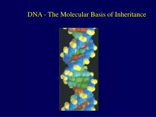
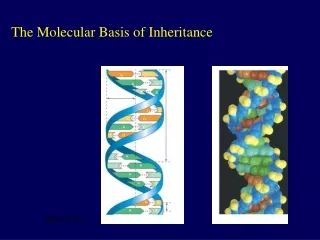
The Molecular Basis of Inheritance. 0. 16. Lecture Presentation by Nicole Tunbridge and Kathleen Fitzpatrick. Life ’ s Operating Instructions. In 1953, James Watson and Francis Crick introduced an elegant double-helical model for the structure of deoxyribonucleic acid, or DNA

E N D
The Molecular Basis of Inheritance 0 16 Lecture Presentation by Nicole Tunbridge and Kathleen Fitzpatrick
Life’s Operating Instructions • In 1953, James Watson and Francis Crick introduced an elegant double-helical model for the structure of deoxyribonucleic acid, or DNA • Hereditary information is encoded in DNA and reproduced in all cells of the body • This DNA program directs the development of biochemical, anatomical, physiological, and(to some extent) behavioral traits
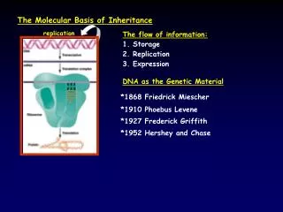
DNA is copied during DNA replication, and cells can repair their DNA
Concept 16.1: DNA is the genetic material • Early in the 20th century, the identification of the molecules of inheritance loomed as a major challenge to biologists
The Search for the Genetic Material: Scientific Inquiry • When T. H. Morgan’s group showed that genes are located on chromosomes, the two components of chromosomes—DNA and protein—became candidates for the genetic material • The role of DNA in heredity was first discoveredby studying bacteria and the viruses thatinfect them
Evidence That DNA Can Transform Bacteria • The discovery of the genetic role of DNA began with research by Frederick Griffith in 1928 • Griffith worked with two strains of a bacterium, one pathogenic and one harmless
When he mixed heat-killed remains of the pathogenic strain with living cells of the harmless strain, some living cells became pathogenic • He called this phenomenon transformation, now defined as a change in genotype and phenotype due to assimilation of foreign DNA
Experiment Living R cells(nonpathogeniccontrol) Mixture of heat-killed S cells andliving R cells Living S cells(pathogeniccontrol) Heat-killed S cells(nonpathogeniccontrol) Figure 16.2 Results Mouse dies Mouse healthy Mouse dies Mouse healthy Living S cells
In 1944, Oswald Avery, Maclyn McCarty, and Colin MacLeod announced that the transforming substance was DNA • Many biologists remained skeptical, mainly because little was known about DNA
Evidence That Viral DNA Can Program Cells • More evidence for DNA as the genetic material came from studies of viruses that infect bacteria • Such viruses, called bacteriophages (or phages), are widely used in molecular genetics research • A virus is DNA (sometimes RNA) enclosed by a protective coat, often simply protein
Phagehead Figure 16.3 DNA Tailsheath Tailfiber Geneticmaterial 100 nm Bacterialcell
In 1952, Alfred Hershey and Martha Chase showed that DNA is the genetic material of a phage known as T2 • They designed an experiment showing that only one of the two components of T2 (DNA or protein) enters an E. coli cell during infection • They concluded that the injected DNA of the phage provides the genetic information
4 4 2 3 1 Experiment Batch 1: Radioactive sulfur (35S) in phage protein Agitation frees outsidephage parts from cells. Centrifuged cellsform a pellet. Labeled phagesinfect cells. Radioactivity(phage protein)found in liquid Radioactiveprotein Figure 16.4 Centrifuge Pellet Batch 2: Radioactive phosphorus (32P) in phage DNA RadioactiveDNA Centrifuge Radioactivity (phageDNA) found in pellet Pellet
Additional Evidence That DNA Is the Genetic Material • It was known that DNA is a polymer of nucleotides, each consisting of a nitrogenous base, a sugar, and a phosphate group • In 1950, Erwin Chargaff reported that DNA composition varies from one species to the next • This evidence of diversity made DNA a more credible candidate for the genetic material
Sugar–phosphatebackbone Nitrogenousbases 5′ end Thymine (T) Figure 16.5 Adenine (A) Cytosine (C) Phosphate Guanine (G) 3′ end Sugar(deoxyribose) DNAnucleotide Nitrogenousbase
Two findings became known as Chargaff’s rules • The base composition of DNA varies between species • In any species the number of A and T bases are equal and the number of G and C bases are equal • The basis for these rules was not understood until the discovery of the double helix
Building a Structural Model of DNA: Scientific Inquiry • After DNA was accepted as the genetic material, the challenge was to determine how its structure accounts for its role in heredity • Maurice Wilkins and Rosalind Franklin were using a technique called X-ray crystallography to study molecular structure • Franklin produced a picture of the DNA molecule using this technique
Figure 16.6 (a) Rosalind Franklin (b) Franklin’s X-ray diffraction photograph of DNA
Franklin’s X-ray crystallographic images of DNA enabled Watson to deduce that DNA was helical • The X-ray images also enabled Watson to deduce the width of the helix and the spacing of the nitrogenous bases • The pattern in the photo suggested that the DNA molecule was made up of two strands, forming a double helix
5′ end C G Hydrogen bond C G 3′ end G C A C T G 3.4 nm Figure 16.7 A T G C C G C G A T 1 nm G C A T C G C G T A G C A 3′ end T A T 0.34 nm 5′ end A T (a) Key features of DNA structure (b) Partial chemical structure (c) Space-filling model
Watson and Crick built models of a double helix to conform to the X-rays and chemistry of DNA • Franklin had concluded that there were two outer sugar-phosphate backbones, with the nitrogenous bases paired in the molecule’s interior • Watson built a model in which the backbones were antiparallel (their subunits run in opposite directions)
At first, Watson and Crick thought the bases paired like with like (A with A, and so on), but such pairings did not result in a uniform width • Instead, pairing a purine with a pyrimidine resulted in a uniform width consistent with the X-ray data
Purine + purine: too wide Figure 16.UN02 Pyrimidine + pyrimidine: too narrow Purine + pyrimidine: widthconsistent with X-ray data
Watson and Crick reasoned that the pairing was more specific, dictated by the base structures • They determined that adenine (A) paired only with thymine (T), and guanine (G) paired only with cytosine (C) • The Watson-Crick model explains Chargaff’s rules: in any organism the amount of A = T, and the amount of G = C
Sugar Figure 16.8 Sugar Thymine (T) Adenine (A) Sugar Sugar Guanine (G) Cytosine (C)
Concept 16.2: Many proteins work together in DNA replication and repair • The relationship between structure and function is manifest in the double helix • Watson and Crick noted that the specific base pairing suggested a possible copying mechanism for genetic material
The Basic Principle: Base Pairing to a Template Strand • Since the two strands of DNA are complementary, each strand acts as a template for building a new strand in replication • In DNA replication, the parent molecule unwinds, and two new daughter strands are built based on base-pairing rules
T A T T T A A A Figure 16.9-3 C G C C C G G G T A T T T A A A T A T T T A A A G C G C G C G C (a) Parental molecule (b) Separation of parental strands into templates (c) Formation of new strands complementary to template strands
Watson and Crick’s semiconservative model of replication predicts that when a double helix replicates, each daughter molecule will have one old strand (derived or “conserved” from the parent molecule) and one newly made strand • Competing models were the conservative model (the two parent strands rejoin) and the dispersive model (each strand is a mix of old and new)
Firstreplication Secondreplication Parent cell (a) Conservative model Figure 16.10 (b) Semiconserva-tive model (c) Dispersive model
Experiments by Matthew Meselson and Franklin Stahl supported the semiconservative model • They labeled the nucleotides of the old strands with a heavy isotope of nitrogen, while any new nucleotides were labeled with a lighter isotope
The first replication produced a band of hybrid DNA, eliminating the conservative model • A second replication produced both light and hybrid DNA, eliminating the dispersive model and supporting the semiconservative model
Experiment 3 2 4 1 Bacteria cultured in medium with 15N(heavy isotope) Bacteria transferred to medium with 14N(lighter isotope) Results DNA samplecentrifugedafter firstreplication DNA samplecentrifugedafter secondreplication Less dense Figure 16.11 More dense Conclusion Predictions: Second replication First replication Conservativemodel Semiconservativemodel Dispersivemodel
DNA Replication: A Closer Look • The copying of DNA is remarkable in its speed and accuracy • More than a dozen enzymes and other proteins participate in DNA replication
Getting Started • Replication begins at particular sites called origins of replication, where the two DNA strands are separated, opening up a replication “bubble” • A eukaryotic chromosome may have hundreds or even thousands of origins of replication • Replication proceeds in both directions from each origin, until the entire molecule is copied
(a) Origin of replication in an E. coli cell (b) Origins of replication in a eukaryoticcell Origin ofreplication Origin of replication Parental (template)strand Eukaryotic chromosome Daughter(new) strand Parental (template)strand Double-strandedDNA molecule Replicationfork Daughter (new) strand Bacterialchromosome Double-strandedDNA molecule Figure 16.12 Replicationbubble Replicationfork Bubble Two daughterDNA molecules Two daughter DNA molecules 0.25 µm 0.5 µm
(b) Origins of replication in a eukaryoticcell Origin of replication Eukaryotic chromosome Parental (template) strand Double-strandedDNA molecule Daughter (new) strand Figure 16.12b Replicationfork Bubble Two daughter DNA molecules 0.25 µm
At the end of each replication bubble is a replication fork, a Y-shaped region wherenew DNA strands are elongating • Helicases are enzymes that untwist the double helix at the replication forks • Single-strand binding proteins bind to and stabilize single-stranded DNA • Topoisomerase corrects “overwinding” ahead of replication forks by breaking, swiveling, and rejoining DNA strands
Primase Topoisomerase 3′ Figure 16.13 RNAprimer 5′ 3′ 5′ Replicationfork 3′ 5′ Helicase Single-strand bindingproteins
DNA polymerases cannot initiate synthesis of a polynucleotide; they can only add nucleotides to an existing 3′ end • The initial nucleotide strand is a short RNA primer
An enzyme called primase can start an RNA chain from scratch and adds RNA nucleotides one at a time using the parental DNA as a template • The primer is short (5–10 nucleotides long), and the 3′ end serves as the starting point for the new DNA strand
Synthesizing a New DNA Strand • Enzymes called DNA polymerases catalyze the elongation of new DNA at a replication fork • Most DNA polymerases require a primer and a DNA template strand • The rate of elongation is about 500 nucleotides per second in bacteria and 50 per second in human cells
Each nucleotide that is added to a growing DNA strand is a nucleoside triphosphate • dATP supplies adenine to DNA and is similar to the ATP of energy metabolism • The difference is in their sugars: dATP has deoxyribose while ATP has ribose • As each monomer of dATP joins the DNA strand, it loses two phosphate groups as a molecule of pyrophosphate
New strand Template strand 5′ 5′ 3′ 3′ Sugar A T T A Base Phosphate Figure 16.14 G G C C DNApoly-merase C C G G OH 3′ A A T T OH P P P P i 3′ C C P Pyro-phosphate OH Nucleotide 5′ 5′ 2 P i
Antiparallel Elongation • The antiparallel structure of the double helix affects replication • DNA polymerases add nucleotides only to the free 3′ end of a growing strand; therefore, a new DNA strand can elongate only in the 5′ to3′ direction
OverviewOrigin of replication 2 1 Leading strand Lagging strand Primer Leading strand Lagging strand Overalldirectionsof replication Figure 16.15 Origin of replication DNA pol III starts tosynthesize leadingstrand. 3′ 5′ RNA primer 5′ 3′ Sliding clamp 3′ DNA pol III 5′ Parental DNA 3′ 5′ 5′ Continuouselongation in the5′ to 3′ direction 3′ 3′ 5′
Origin of replication DNA pol III starts tosynthesize leadingstrand. 1 3′ 5′ RNA primer 5′ 3′ Figure 16.15b Sliding clamp 3′ DNA pol III 5′ Parental DNA 3′ 5′ 5′ Continuouselongation in the5′ to 3′ direction 2 3′ 3′ 5′
Along one template strand of DNA, the DNA polymerase synthesizes a leading strand continuously, moving toward the replication fork
To elongate the other new strand, called the lagging strand, DNA polymerase must work in the direction away from the replication fork • The lagging strand is synthesized as a series of segments called Okazaki fragments, which are joined together by DNA ligase