Download
1 / 83
830 likes | 1.11k Views
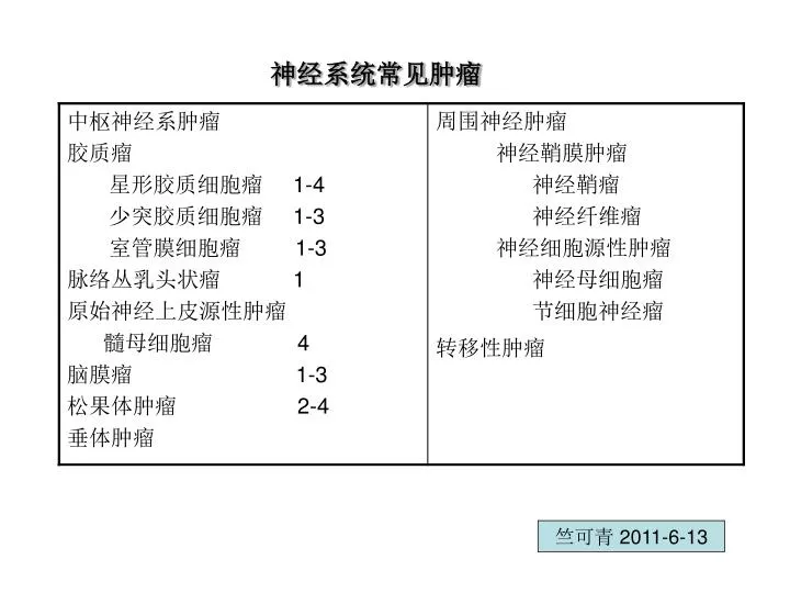
神经系统常见肿瘤. 竺可青 2011-6-13. 中枢神经系统肿瘤. 中枢神经系统肿瘤 发病率: 10-12/10 万 包括起源于脑、脊髓或脑膜的原发性和转移性肿瘤两大类。 原发性肿瘤中, 40% 为胶质瘤, 15% 为脑膜瘤,约 8% 为听神经瘤。 转移性肿瘤以转移性肺癌为多见。 儿童颅内恶性肿瘤仅次于白血病,常见为胶质瘤和髓母细胞瘤。 共同特征:局部神经症状,颅内高压症状,分化良好的肿瘤也可因压迫重要的部位致死,分化很差的肿瘤也很少颅外转移。. WHO 中枢神经系统肿瘤病理 (7 类 ). 神经上皮组织肿瘤( Neuroepithelial tumours)

E N D
神经系统常见肿瘤 竺可青 2011-6-13
中枢神经系统肿瘤 • 中枢神经系统肿瘤发病率:10-12/10万 • 包括起源于脑、脊髓或脑膜的原发性和转移性肿瘤两大类。 • 原发性肿瘤中,40%为胶质瘤,15%为脑膜瘤,约8%为听神经瘤。 • 转移性肿瘤以转移性肺癌为多见。 • 儿童颅内恶性肿瘤仅次于白血病,常见为胶质瘤和髓母细胞瘤。 • 共同特征:局部神经症状,颅内高压症状,分化良好的肿瘤也可因压迫重要的部位致死,分化很差的肿瘤也很少颅外转移。
WHO中枢神经系统肿瘤病理(7类) 神经上皮组织肿瘤(Neuroepithelial tumours) 脑膜肿瘤(Meningeal tumours) 颅内和外周神经肿瘤(Tumours of cranial and peripheral tumours) 淋巴造血系统肿瘤(Tumours of the heamopoietic system) 生殖细胞肿瘤(Germ cell tumours) 鞍区肿瘤(Tumours of the sellar region) 转移性肿瘤(Metastatic tumours of the CNS)
神经上皮组织肿瘤(10类) 星形细胞肿瘤(Astrocytic tumours) 少突胶质细胞肿瘤(Oligodendroglial tumours) 混合性胶质瘤(Mixed gliomas) 室管膜肿瘤(Ependymal tumours) 脉络丛肿瘤(Choroid plexus tumours) 起源不定的胶质肿瘤(Neuroepithelial tumours of uncertain origin) 神经元和混合性神经元-胶质肿瘤(Neuronal and mixed neuronal-glial tumours) 神经母细胞瘤 (Neuroblastoma) 松果体实质肿瘤(Pineal parenchymal tumours) 胚胎性肿瘤(Embryonal tumours)
星形胶质细胞瘤(astrocytic tumours) 颅内最常见的胶质瘤 G:境界不清的灰白色浸润性肿瘤,或硬,或软,或呈胶冻状, 可形成含有清亮液体的囊腔 。 M:星形细胞瘤 (纤维型 原浆型 肥胖型 混合细胞型 ) 间变型星形细胞瘤 胶质母细胞瘤 星形细胞瘤胞质表达胶质纤维酸性蛋白(GFAP*),S-100和波形蛋白。
纤维型星形细胞瘤(fibrillary astrocytoma) 最常见,瘤细胞分化好,但呈浸润型生长。瘤细胞间可见红染的原纤维性背景。 原浆型星形胶质细胞瘤(protoplasmic astrocytoma) 为少见类型,瘤细胞体小,胞突少而短。 肥胖型星形胶质细胞瘤(gemistocytic astrocytoma) 瘤细胞体大,胞质丰富。 间变性星形细胞瘤(anaplastic astrocytoma) 瘤细胞密度增加,多形性,核异态及核分裂像等,为恶性肿瘤。此型经常表现为快速进展,最终转变为胶质母细胞瘤。
胶质母细胞瘤 (glioblastoma multiforme, GBM WHO4级) 镜下:细胞密集,异型性明显,可见怪异单核或多核巨细胞。 出现出血坏死。发展迅速,预后极差。 毛发细胞型星形胶质细胞瘤(pilocytic astrocytoma WHO I级) 多发生在儿童、青少年,生长极为缓慢。 多形性黄色星形细胞瘤(Pleomorphic xanthoastrocytoma WHOII级) 好发儿童和年轻人,大脑半球表面(颞叶)
St.anne/mayo分级系统(核的异型、分裂象、内皮细胞增生/或坏死)St.anne/mayo分级系统(核的异型、分裂象、内皮细胞增生/或坏死) 星形细胞瘤II级 1个标准 星形细胞瘤III级 2个标准 星形细胞瘤IV级 3个标准
Specific structure of intracranial tumor 实质的特征性结构 间质的特征性结构 继发性变化:神经元周围的卫星现象 血管周围肿瘤细胞聚集 软脑膜下肿瘤细胞聚集
颅内肿瘤症状 • 局部压迫神经症状 • 颅压升高症状
一、中枢神经肿瘤 (一)胶质瘤(glioma) • 良恶性的相对性,无包膜,均呈浸润性生长,间变的界限反而清楚。 • 局部浸润 • 转移:① 脑脊液转移为常见方式 ② 颅外转移极少见
GLIOMAS • Gliomas, derived from glial cells, include • astrocytomas, • oligodendrogliomas, and • ependymomas • Among the diffuse fibrillary astrocytomas, tumors can be • well differentiated (astrocytoma) or • less differentiated (higher-grade), ranging from • anaplastic astrocytoma to • glioblastoma.
WHO 1-4 grade • Pilocytic astrocytoma 1 • Well-differentiated fibrillary astrocytomas 1 • Gemistocytic astrocytoma 2 • Anaplastic astrocytomas 3 • Glioblastoma (glioblastoma multiforme) 4
1.星形胶质细胞瘤(astrocytoma) • 约占颅内肿瘤的30%,占胶质瘤的70% • 男性多见 • 肉眼:数厘米→巨大 分化好→界不清;分化差→界清 灰白色,质地或软或硬或呈胶冻状 脑原有的结构受压变形
镜下:形态多样, 纤维型 良 1 原浆型 肥胖型——交界 2 间变性星形胶质细胞瘤——恶性 3 胶质母细胞瘤——恶性 4 • 特点:细胞间变,密度大,异型性多,核深染,毛细血管内皮细胞增生。
Diffuse fibrillary astrocytoma. 弥漫性纤维性星形细胞瘤
Diffuse fibrillary astrocytoma. An immunoperoxidase preparation demonstrates labeling of tumor cell bodies and processes by a monoclonal antibody to glial fibrillary acidic protein.
高度恶性星形胶质细胞瘤,又称多形性胶质母细胞瘤(WHO 4级)。 • 好发额、颞叶白质。 • 浸润广可至对侧。 • 镜下:细胞密集,异型性明显。 怪异核,多核瘤巨细胞。 出血坏死明显——区别于间变型的特征 毛细血管及内皮细胞增生,肿大 发展迅速,预后差,2年内死亡
Well-differentiated astrocytoma. The right frontal tumor has expanded gyri, which led to flattening.
Expanded white matter of the left cerebral hemisphere and thickened corpus callosum and fornices
Computed tomographic (CT) scan of a large tumor in the cerebral hemisphere showing signal enhancement with contrast material and pronounced peritumoral edema
Glioblastoma multiforme appearing as a necrotic, hemorrhagic, infiltrating mass.
Glioblastoma. Foci of necrosis with pseudopalisading of malignant nuclei
(c) Small cell 小细胞型 survival 6.6-11.5 mo, exocranial metastasis
毛细胞型星形胶质细胞瘤 Pilocytic astrocytoma • 发生于儿童,青少年 • 生长缓慢,带瘤存活达40年 • 常位于小脑,第四脑室底部,第三脑室丘脑,视神经 • 特点为多极性的肿瘤细胞两端长出纤细毛发状突起 • 预后好 • GFAP为胶质瘤的标志 • 原癌基因sis过度表达
Pilocytic astrocytoma in the cerebellum with a nodule of tumor in a cyst
2.少突胶质细胞瘤 oligodendroglioma • 占颅内神经上皮性肿瘤5% • 60-40岁成人 • 部位: 大脑半球皮质浅层,额叶多见 灰红色边界清楚的球形肿瘤 囊性变,出血,钙化常见。
瘤细胞大小均匀,形态单一,弥漫排列。 • 核深、浆空呈空晕状。 • 间质富有血管,不同程度内皮细胞增生。 • 20%有钙化。 • 生长慢——达10-30年。 • 临床多为癫痫和局部性瘫痪。 • 少数生长迅速,预后不佳。
Oligodendroglioma. Uniform, round nuclei and clear perinuclear halos (artefacts of delayed fixation) typify well-differentiated oligodendrogliomas
3.室管膜细胞瘤 • 多为儿童、青少年。 • 占5%-6% • 多见于第四脑室,次为侧脑室,三脑室和导水管。 • 呈球形、分叶或乳头状,界清,膨胀性生长于脑室内,切面灰白均匀或颗粒状。 • 细胞大小一致,梭形或胡萝卜状。 • 核膜清,有核仁胞浆丰富。 • 有菊形团及假菊形团结构,也可呈乳头状结构。 • PTAH染色,可见纤毛体。
Ependymoma. Tumor growing into the fourth ventricle, distorting, compressing, and infiltrating surrounding structures.
(二)髓母细胞瘤 • 多见于15岁以下儿童 男>女 • 来源于小脑蚓部原始神经上皮细胞或小脑皮质的胚胎性外颗粒层细胞。 • 瘤组织灰红色肉状。 • 细胞密集呈圆椭圆或胡萝卜形。 • 胞浆少而边界不清。 • 围绕纤细嗜银性纤维呈放射状排列——菊形团。 • 高度恶性,预后差。
Medulloblastoma. CT scan showing a contrast-enhancing midline lesion in the posterior fossa.
Sagittal section of brain showing medulloblastoma destroying the superior midline cerebellum.
二、脑膜瘤(menignioma) • 来源脑膜,生长缓慢,大多为良性。 • 40-50岁女性多见,颅内脑膜瘤比脊髓多2倍。有14%无症状脑膜瘤 • 好发于蛛网膜颗粒所在部位。 • 球形,分叶或不规则状。 • 质实,硬,边界清,周围脑组织受压。 • 切面呈灰白,颗粒状或条索旋涡状,有时有砂砾样。
MENINGIOMAS • Atypical meningiomas (WHO grade II/IV) • Anaplastic (malignant) meningioma (WHO grade III/IV)