Download
1 / 39
410 likes | 476 Views
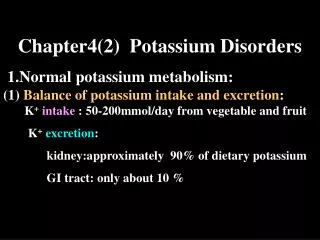
Explore the intricate mechanisms of normal potassium balance, factors influencing potassium levels, and the etiology, consequences, and treatment of potassium disorders in the body.

E N D
Normal Potassium Balance 100 mEq K+ RBC ~250 Extracellular fluid 65 mEq Muscle ~2500 Liver ~250 Bone ~500 90-95 mEq 5-10 mEq
Extra-renal K+ Homeostasis: Shift/Redistribution • 4 major factors: - • Hormones • Acid base status • Plasma tonicity • Plasma [K+]
Mineralocorticoids (Aldosterone):- • Produced by ZonaeGlomerulosa of the Adrenal cortex :- • Release stimulated by:- • Activation of Renin/Angiotensin system:- • Volume sensors, JGA etc. • Direct effect of K+ on adrenal. • Effects of Aldosterone:- • Exchange of Na+ for K+ or H+ with net loss of K+ and gain of sodium.
Mineralocorticoids (Aldosterone):- • Sites of action: • Distal renal tubule • Colon • Sweat Glands • In renal failure, the colon is an important site for K+ regulation • Cortisol has mineralocorticoid activity
Acid/Base • Metabolic Acidosis [K+]: - • Inhibition of renal tubular K+ secretion • Shift of K+ fromICF to ECF • Metabolic Alkalosis [K+]
ECF [K+] depends upon: • Redistribution: ECF and ICF • Hormones • Acid base status • Plasma tonicity • Plasma [K+] Output Kidney Sweat glands Gut (colon) Intake
Proximal Tubule (60-70%) Ascending Loop of Henle (20-30%) Distal tubule: 10% remaining 100% K+ Reabsorbed 600 mmol/day K+ Filtered Distal Tubule = Fine tuning. Aldosterone K+/H+ for Na+ 100 mmol/day K+ excreted (10-20%) Renal Handling of K+ Glomerulus Renal artery
K+ <5.0 mmol/L K+ <5.0 mmol/L Sensitive to K+ Aldosterone, pH Aldosterone Sensitive K+ Cl- Principal Cells Tubular Lumen Peritubular Capillary K+ 100mmol/L Na+ K+ Na/K ATPase -50 mV Na+ 3 Na+ for 2 K+
Factors favouring K+ secretion • Increased Na+ reabsorption • Exchange • Increased intra-cellular K+ • Concentration gradient • Decreased H+ secretion. • Aldosterone • Electrochemical gradient.
Increased fluid delivery to lumen causes increased K+ excretion: - • Wash out. • diuretics • poorly absorbed anions • osmotic diuresis
Electrochemical Gradient: - • Distal tubule actively secretes H+ • K+ and H+ are neutralising the same electrical gradient Acidosis Causing Hyper-kalaemia
Etiology of Hyperkalemia • Increased intake • Movement from cells into extracellular fluid • Decreased urinary excretion • Combinations of above
Movement from cells into ECF Pseudohyperkalemia Movement of K+ out of cells during or after blood drawing Hemolysis Fist clenching (local exercise effect) Marked leukocytosis Metabolic acidosis – non organic acids Insulin deficiency Hyperosmolality Beta blockade
Movement from cells into extracellular fluid • Tissue catabolism • Trauma • Cytotoxic agents (TLS) • Severe hypothermia • Severe exercise • Digitalis overdose • Hyperkalemic periodic paralysis • Post cardiac surgery • Succinylcholine
Hypoaldosteronism • Decreased activity of renin-angiotensin system • Hyporeninemic hypoaldosteronism (T4 RTA) • NSAIDS, ACE-I, CSA • Primary decrease in adrenal synthesis • Adrenal insufficiency, CAH, heparin • Aldosterone resistance • K+sparing diuretics, trimethoprim, CSA • pseudohypoaldosteronism
Hyperkalaemia • Reduced excretion • Renal Failure • Mineralocorticoid deficiency (Addison's) • Tubular defects • Drugs • Potassium sparing diuretics (spiranolactone) • Inappropriate use of K supplements with above Factitious • Improper collection • Haematological disorders Increased input • Oral • IV therapy Altered distribution • Acidemia • insulin deficiency • Crush Injury or haemolysis
Hyperkalaemia Most usual causes • Decreased renal function • Redistribution secondary to acidosis Consequences of Hyperkalaemia • Neuromuscular - Weakness, parasthesia, paralysis • Gastrointestinal - Nausea, vomiting, pain, ileus • Cardiovascular - Conduction defects, arrhythmias, Cardiac arrest
S&S of Hyperkalemia • Muscle weakness – decr magnitude of resting MP • Cardiac toxicity Enhanced by : • Hypocalcemia • Acidemia • Hypomagnesemia • Rapid onset
Treatment of Acute Hyperkalemia • Assess urgency • Stabilize myocardium: Ca-Gluconate • Redistribute K+ from ECF to ICF: Insulin/D50, Albuterol (high dose), NaHCO3. • Remove K+ from body: kayexalate,diuretics, and dialysis
Treatment of chronic hyperkalemia • Limit intake! • 2 g/day • OTC that contain K+ • Avoid drugs that induce (NSAIDs!) • Increase K excretion • Diuretics, avoid constipation, +/- kayexalate
Etiology of Hypokalemia 1) Decreased net intake 2) Increased entry into cells 3) Increased GI losses 4) Increased urinary losses 5) Increased sweat losses 6) Other losses (dialysis)
Decreased net intake • Normal 100 mEq/d (40-120) • Renal excretion as low as 5–20 mEq/d • Thus, decreased intake rare cause, but increases susceptibility to hypokalemia • Clay ingestion can bind dietary K+ in the gut reducing absorption.
Increased Entry into Cells • Elevated extracellular pH • H+ released from cellular buffers • Increased availability of insulin • Elevated beta-adrenergic activity
Increased Entry into Cells • Periodic paralysis: • Familial, autosomal dominant, onset up to 30 yo, severe. • Acquired with thyrotoxicosis (Asian men) • Episodes precipitated by: • Rest post-exercise • Carbohydrate meal • Stress, cold • Administration of insulin or epinephrine • Sudden movement of K+ into cells, acute decrease in [K+] • Plasma [K+] is normal between attacks
Increased GI losses • Vomiting • Diarrhea • Intestinal fistulas • Tube drainage • Loss of colonic secretions • Villous adenoma • Laxative abuse
Increased urinary losses • Diuretics • Increased flow to K+ secretory sites • Enhanced secretion of aldosterone • Bartter’s or Gitelman’s Syndrome • Mineralocorticoid excess. • Reabsorption of Na+ in presence of nonreabsorbable anion. • RTAs.
Consequences of Hypokalaemia • Skeletal muscle - Weakness, paralysis • Gastrointestinal - Paralytic Ileus • Kidney - Impaired concentrating ability, Tubular defects • Cardiac - Conduction defects, arrhythmias, Digoxin toxicity
Hypokalemia and Hypertension Urine K+ excretion > 30mEq/d < 25 mEq/d Plasma renin activity GI losses Prior diuretic Rx Low High or normal Plasma aldosterone High Low Continued diuretic use Renovascular HTN Malignant HTN Renin-secreting tumor Cushing’s Syndrome Primary Hyperaldosteronism Licorice Liddle’s SAME Other Mineralo- corticoid Adrenal vein aldo +/- CT Adenoma: Do surgery Hyperplasia: K+ sparing diuretic
Role of cortisol • Cortisol binds to mineralocorticoid receptor with equal avidity to aldosterone • Conversion to cortisone by 11-hydroxysteroid dehydrogenase prevents mineralocorticoid activity • Cortisol acts as mineralocorticoid with: • Excess cortisol production: Cushing’s • Impairment of 11- : real licorice, syndrome of AME
Work-up of hypokalemia • H&P • Diarrhea • Vomiting • Diuretics • Periodic paralysis • Offending drugs • Acid-base balance • Urine potassium • >25-30 meq/ indicates renal losses • < 15 meq/L on spot usually rules out urine losses
Hypokalaemia: Treatment Not urgent UNLESS complications • Oral is preferable to IV therapy • Deficits: • K = 3 mmol/L ~ 300 mmol • K = 2 mmol/L ~ 600 mmol Oral treatment • Normal intake + 60 mmol/day • Slow K …. 8 mmol. • I.V. treatment • 10 mmol/hr Maximum!
55 yo lady with DM >10 years associated with DM nephropathy and retinopathy, HTN >10 years, CKD III with baseline Cr around 1.8-2.0 presented for regular clinic visit. Na 138, K 5.7, HCO3 18, Cl112, Cr 1.8. Urine pH 5.0. ABGs showed 7.32/34/18. What is the most likely diagnosis? Diarrhea induced metabolic acidosis. Methanol overdose. Ethylene Glycol overdose RTA 4 Distal RTA
70 yo man presented to the ER c/o fever, chills and SOB for one week. CXR showed bilateral pneumonia. BP is 100/60 (baseline around 140s-150s/80s-90s). Lab showed Cr of 5.0. K 7.0. EKG showed hyper-acute T waves. What is the best next step? • IV calcium gluconate. • PO kayexalate. • IV furosemide • Emergent HD catheter and dialysis. • Call the nephrology team and tell them to come and take care of their patient since you are busy and have other patients to see in the ER.
58 year old lady presented to the ER with weakness. Na 136. K 3.1. Cl 110. CO2 15. ABGs showed pH 7.28. PaCO2 30. HCO3 16. Urine: Na 10 meq/l, K 8 meq/l, Cl- 40 meq/l. What is the most likely diagnosis? • DKA • Lactic acidosis • Severe vomiting • Severe diarrhea • RTA
24 year old lady presented to the ER with weakness. BP 85/45. BMI 35. Na 138. K 2.8. CO2 34. Mg 1.2. ABGs 7.49/45/34. She has been trying hardly to lose weight but denies laxatives or diuretics use. What is the best next step to reach a most likely diagnosis? • Given low serum Mg along with other lab values she most likely has Gitelman syndrome. • Given low serum Mg along with other lab values she most likely has Bartter syndrome. • She is most likely abusing laxatives. Ask more questions about laxatives abuse. • Send a blood and urine sample for diuretics screen. • She most likely had anorexia nervosa.